Recurrent VT with ICD shocks
Raja Selvaraj JIPMER
Clinical presentation
Presentation (2012, elsewhere)
- 45 year old male
- Dyspnea / fatigue
- LV dysfunction - EF 40%
- CAG normal
2016
- Palpitations
- ? Documented tachycardia
- What is appropriate management if sustained VT ?
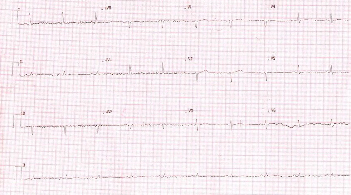
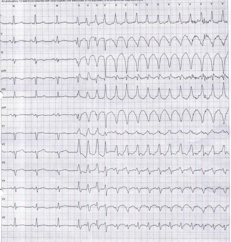
ECG
NICM / sustained VT
- Beta blockers
- Amiodarone
- ICD
- CRTD
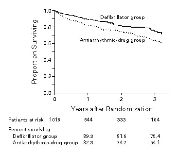
AVID
Further course
- Single chamber ICD implanted
- Multiple shocks in Jan 2017
- ICD interrogation - appropriate therapy for VT
- Already on Amiodarone 200 mg OD
- Opinion in different hospitals
ICD, VT on Amiodarone - what would you do?
- Increase dose of amiodarone
- Add Mexilitene
- RF ablation
- Check ICD
Further course
- May 2017 - Multiple NSVT
- Increased Amio 300 mg OD
- Jan 2018 - 4 shocks
- Came asking for ICD to be turned off
- Device interrogation - 180 episodes of NSVT since Jan 2018
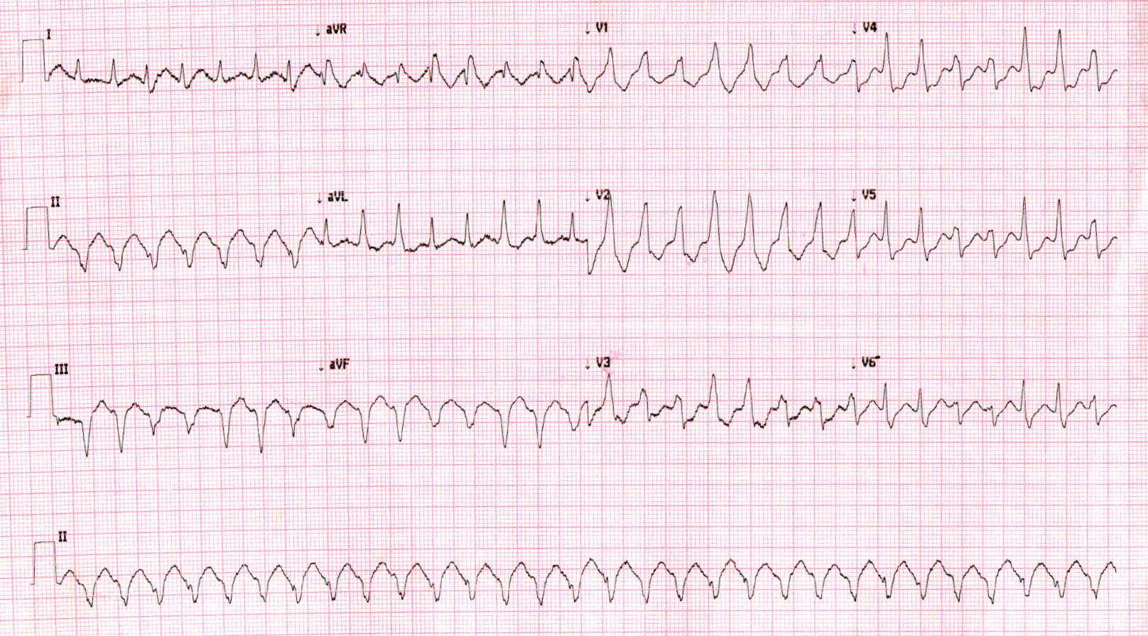
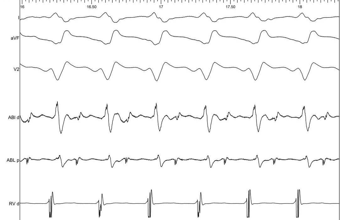
ECG - Where is origin ?
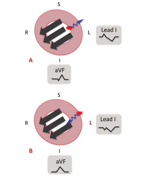
ECG clues for epicardial origin
- Interval criteria
- Pseudo delta
- Intrinsicoid deflection time
- Shortest RS
- Maximum deflection index
- Morphologic criteria
- q wave in lead I
- q wave in inferior leads
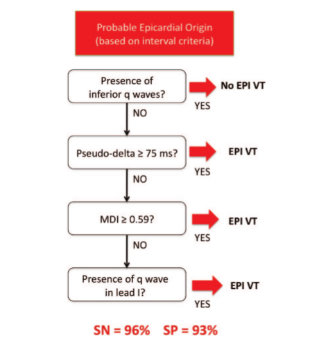
ECG Criteria to Identify Epicardial Ventricular Tachycardia in Nonischemic Cardiomyopathy. Ermengol Vallès, Victor Bazan and Francis E. Marchlinski. Circulation: Arrhythmia and Electrophysiology. 2010;3:63-71
Imaging
- Echo: LVEF 40-45%
- MRI: Scar in IVS / lateral LV
- Mapping and ablation plan ?
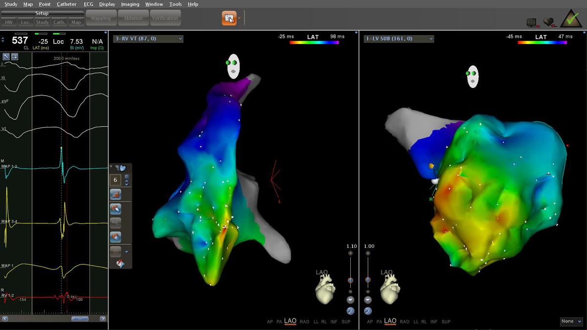
Endocardial mapping
Initial approach
- Attempted percutaneous pericardial access
- Unable to access pericardial space
- LV endocardial map
LV voltage map (RV pacing)
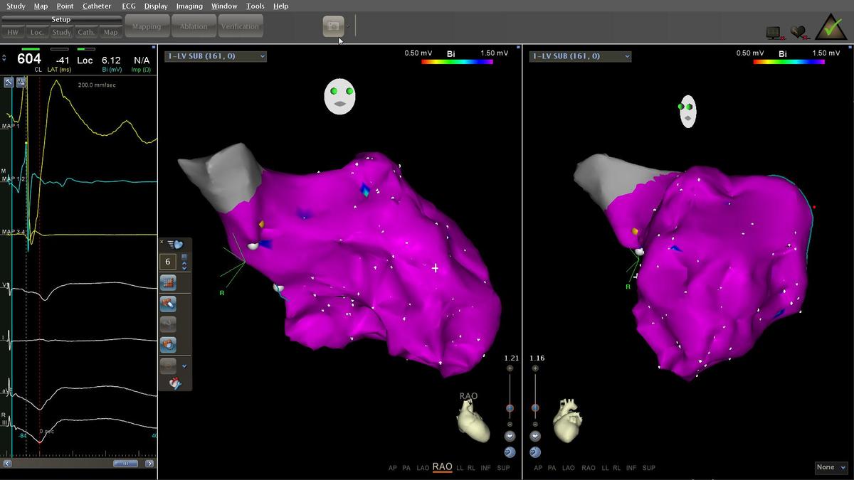
What to do?
- Unipolar voltage
- Induce VT
- Empiric ablation
Endocardial unipolar voltage predicts epicardial bipolar voltage
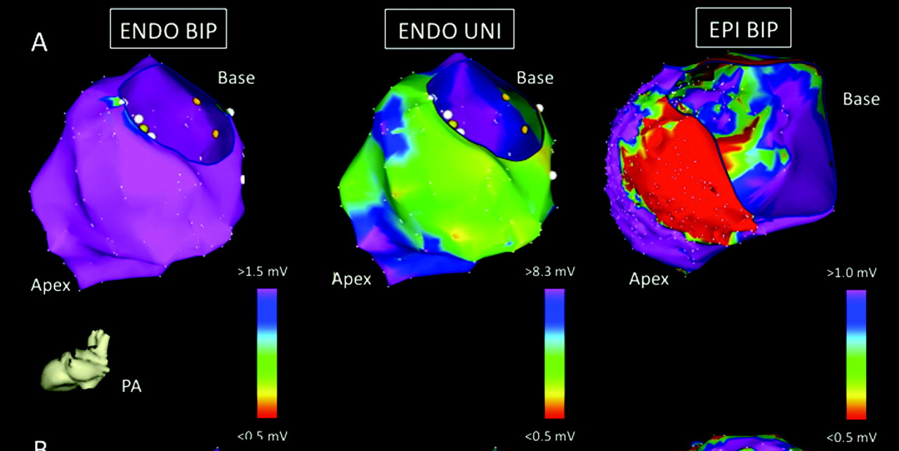
- Hutchinson … Marchlinski. Endocardial unipolar voltage mapping to detect epicardial ventricular tachycardia substrate in patients with nonischemic left ventricular cardiomyopathy. Circ Arrhythm Electrophysiol 2011;4:49
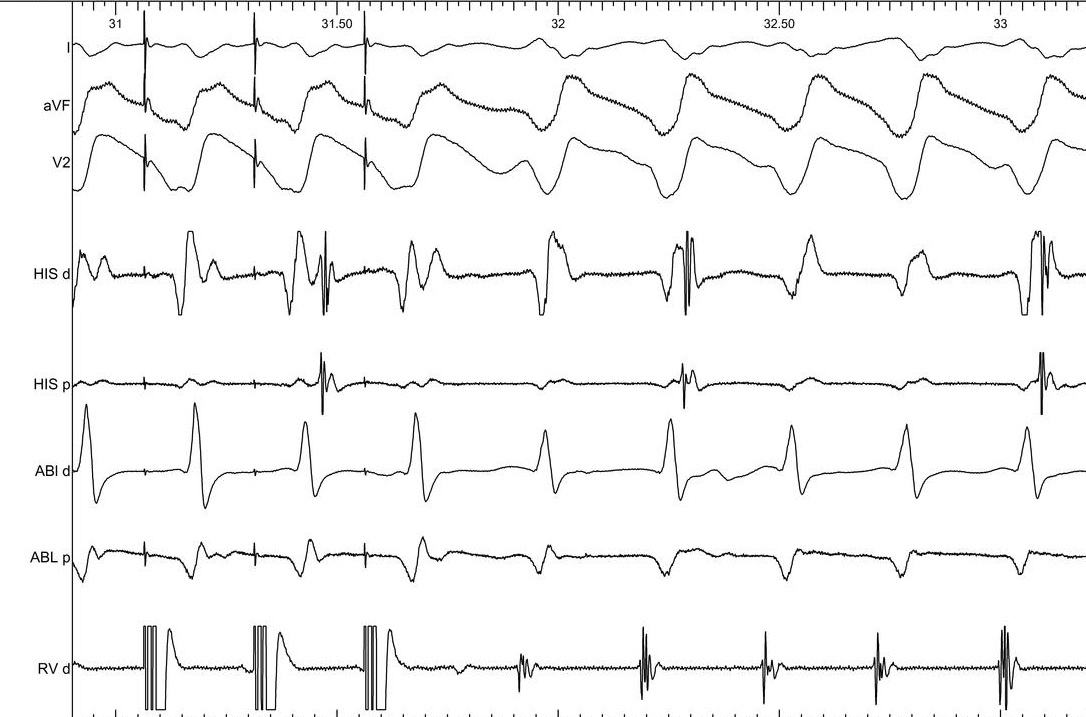
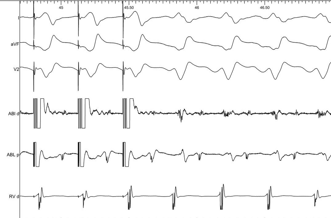
Tachy induction - what is interpretation ?
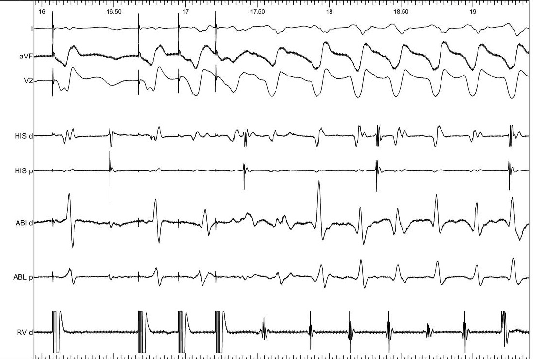
VT - What is the V-H relationship ?
VT map - LV and RV
RV entrainment
Epicardial mapping
Subsequent course
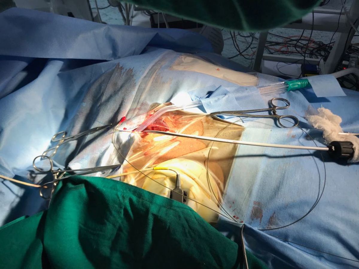
- Subxiphoid window
- Wire in pericardial space
- Agilis epi sheath
Wire in pericardial space
Setup
Epicardial map - Rt Lat
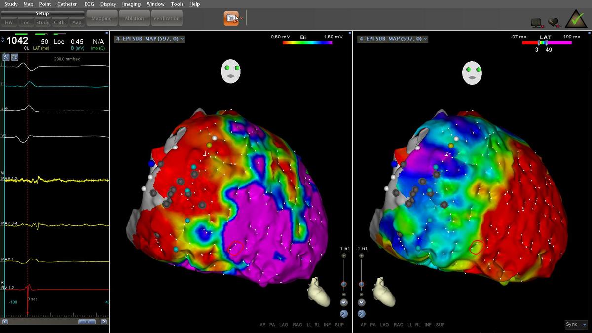
Epicardial low voltage - fat / scar
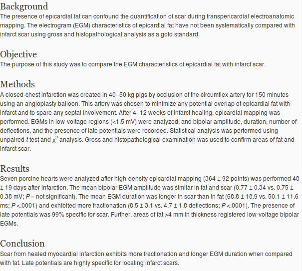
Distinguishing epicardial fat from scar: Analysis of electrograms using high-density electroanatomic mapping in a novel porcine infarct model. Shivkumar. Heart Rhythm 2010
Epicardial map - Lt Lat
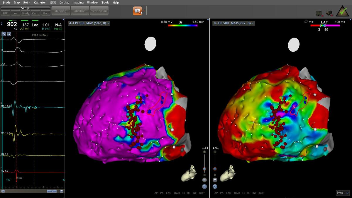
Substrate in inferolateral LV
- what are potential innocent bystanders ?
- Phrenic nerve
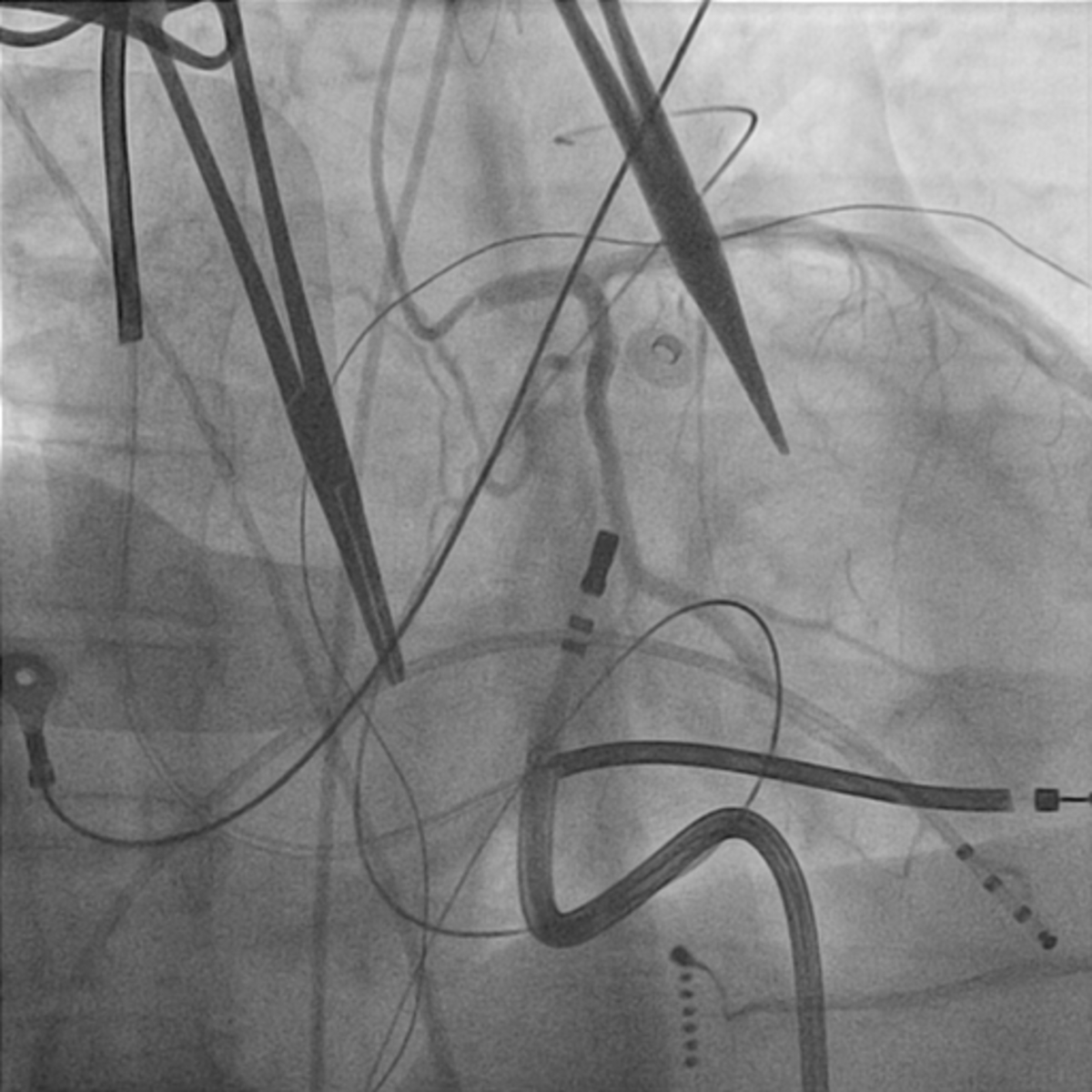
- Coronary arteries
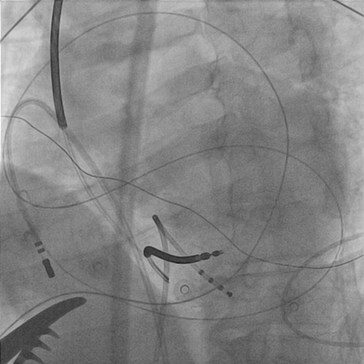
LCA angiogram
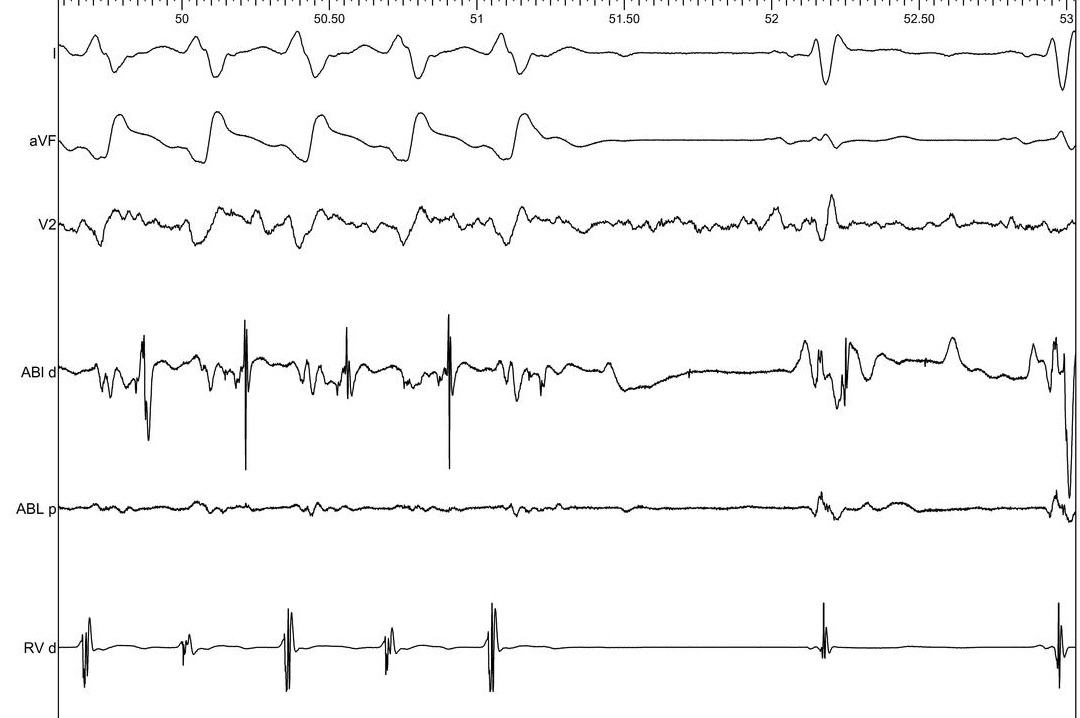
Signal during VT - where is the catheter
Where is the catheter ?
- Outer loop
- Isthmus entrance
- Isthmus exit
- Isthmus center
- Dont know / cant tell
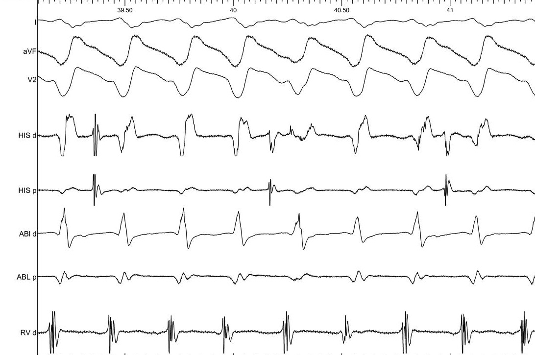
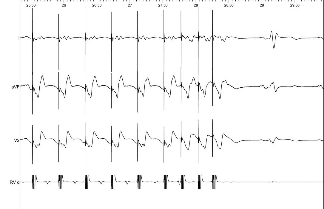
Entrainment
Where is the catheter ?
- Outer loop
- Isthmus entrance
- Isthmus exit
- Isthmus center
- Dont know / cant tell
Further course
- Started ablating
- Terminated with ablation further lower
- Subsequent ablation to eliminate late potentials
Post ablation VT induction
Learning points
- Identification of epicardial VT on ECG
- Unipolar voltage to identify epicardial scar
- Sub-xiphoid window
- Pitfalls / perils in epicardial mapping