
Introduction
What is electroanatomic mapping ?
- Anatomical information collected with electrical information
- In the same system
What does it do
Memory aid
Visualization tool for electrical information
- Any aspect of electrical information that can be quantified
- Display over the anatomical map
Quantification of electrical information
- Bipolar voltage
- Activation time in relation to a fixed time reference
- Unipolar voltage
Catheter visualization
- Continuous
- Non-fluoroscopic
Visualize geometry of chamber
- Collect only intermittently
- Or collect continuously (FAM)
- But how does the system know where the walls (shell) are ?
Image integration from other modalities
- CT / MRI
- Rotational angio
- Ultrasound
What it does not do
- Automatically identify the mechanism of tachycardia
- Automatically identify the site of ablation
- Free operator from need to exercise judgement
The "self driving" car

When allowed to drive by itself

Why ? - or why not just conventional mapping ?
Ablate arrhythmias that we (almost) cannot otherwise
- Scar based VT
- Atypical atrial flutter
Reduce fluoroscopy significantly
- Atrial fibrillation
- Typical atrial flutter
Reduce fluoroscopy marginally (balance with costs)
- AVNRT
- AVRT
- Idiopathic ventricular tachycardia
- Focal atrial tachycardia
Record ablation points
- Helpful for linear ablation
- new technologies incorporating various parameters to color code ablation points
Other benefits
- Improved success ?
- Reduce complications ?
Evidence
- RCT - 102 patients undergoing ablation for various arrhythmias in 2004
- CARTO vs conventional
- Success and duration same
- Reduced radiation
- Increased cost
- similar results in other studies (2,3)
- Sporton S … Schilling R. Electroanatomic Versus Fluoroscopic Mapping for Catheter Ablation Procedures: A Prospective Randomized Study. JCE 2004;15:310-315
- Khongphatthanayothin … Nademanee. Nonfluoroscopic Three‐Dimensional Mapping for Arrhythmia Ablation: Tool or Toy? JCE 2000;11:239-243
- Ealey M et al. Radiofrequency ablation of arrhythmias guided by non-fluoroscopic catheter location: a prospective randomized trial. EHJ 2006;27:1223–1229,
Technology
Magnet based
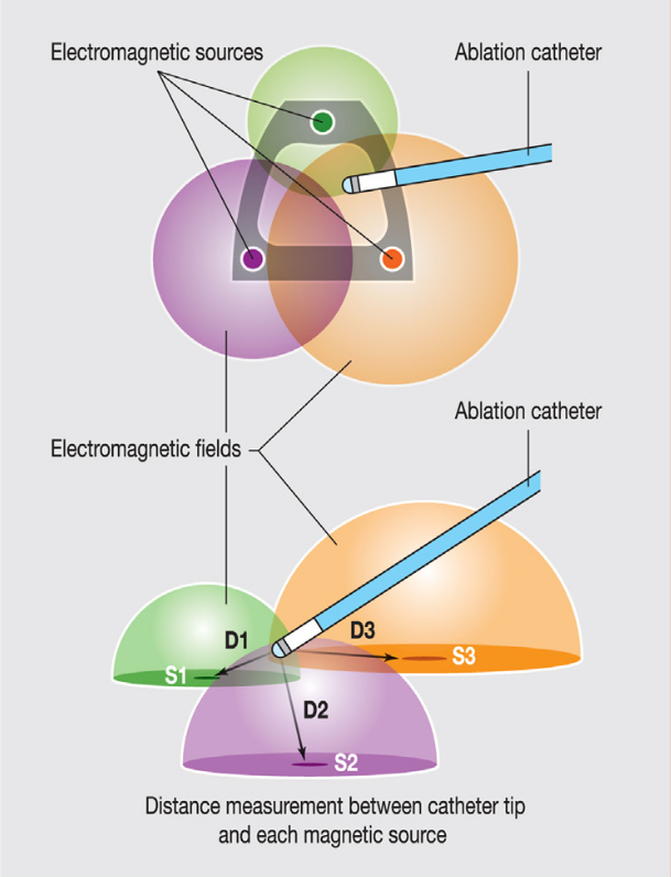
Magnet based
- pros
- Accurate
- cons
- Needs special catheter with magnetic sensor at tip
Impedance based
- Low amplitude, high frequency current
- Six patches in three orthogonal directions
Impedance based
- pros
- Can be used with any catheter / any electrode / anything like an electrode
- cons
- Impedance less reliable, especially over time
Systems
- CARTO
- NavX
- Rhythmia
Point by point
- pros
- Every point validated
- Ablation catheter itself
- cons
- Takes time
- False annotations may affect more
Multi point
- pros
- faster
- more points
- cons
- Cannot check each point
- Need separate catheter for ablation
- Few false annotations may have less weight
Technical aspects in practice
Types of maps
- Anatomical
- Voltage
- Activation / propagation
- Others
Anatomical maps
- Only geometry acquired
- Atrial fibrillation
- AVNRT
Voltage maps
- Iso-voltage maps - EGM amplitude
- Cut-offs derived from small set of patients
- Varies based on catheter
- Normal > 1.5 mV, abnormal < 1.5, scar < 0.5, dense scar ?
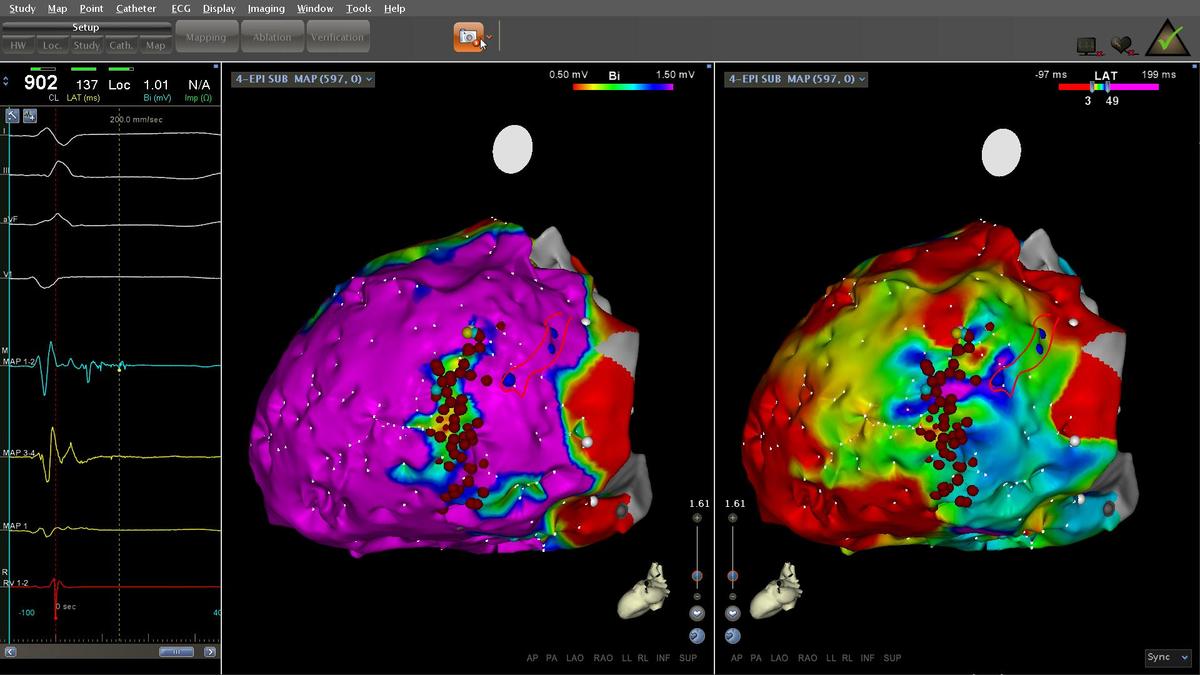
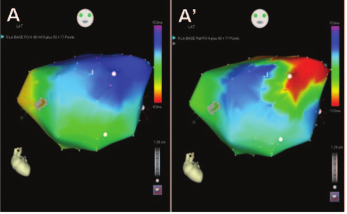
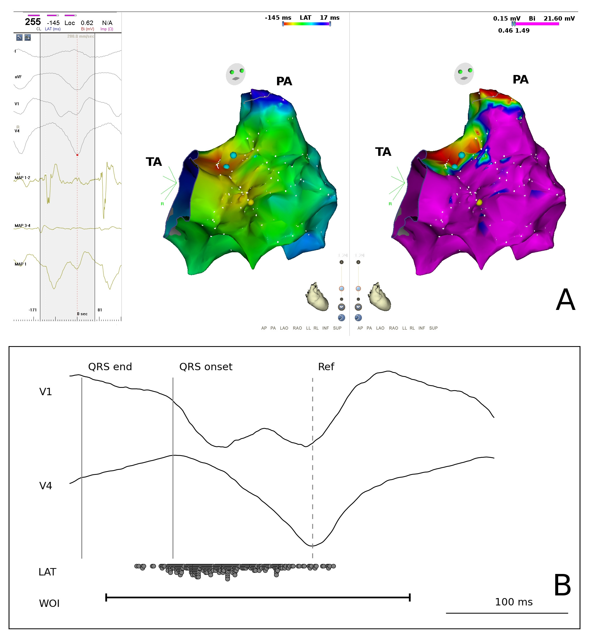
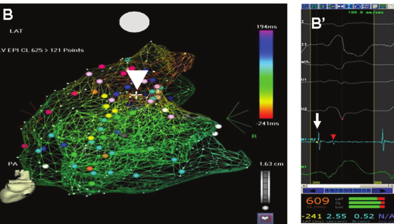
Voltage map
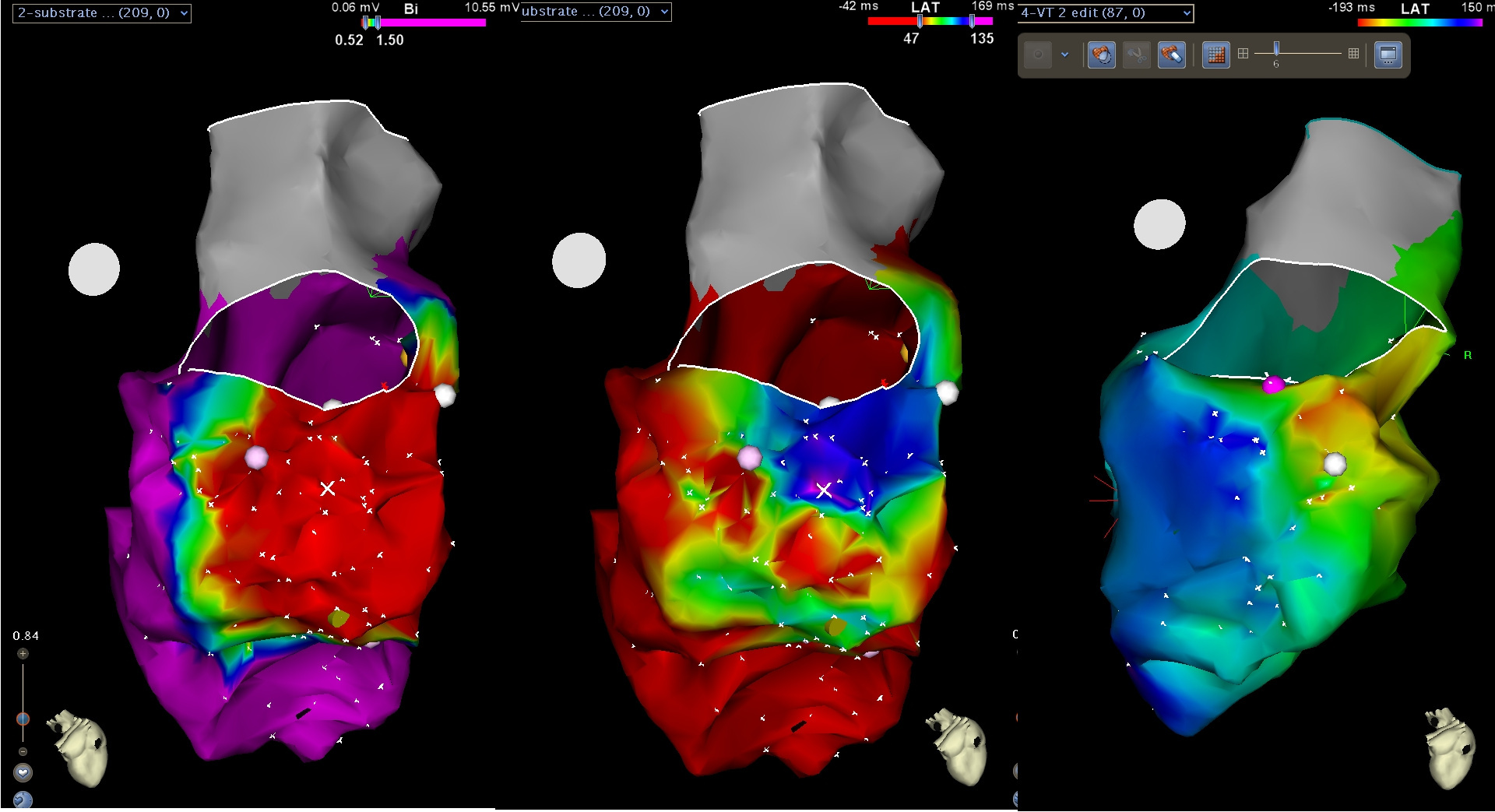
voltage map in epicardium
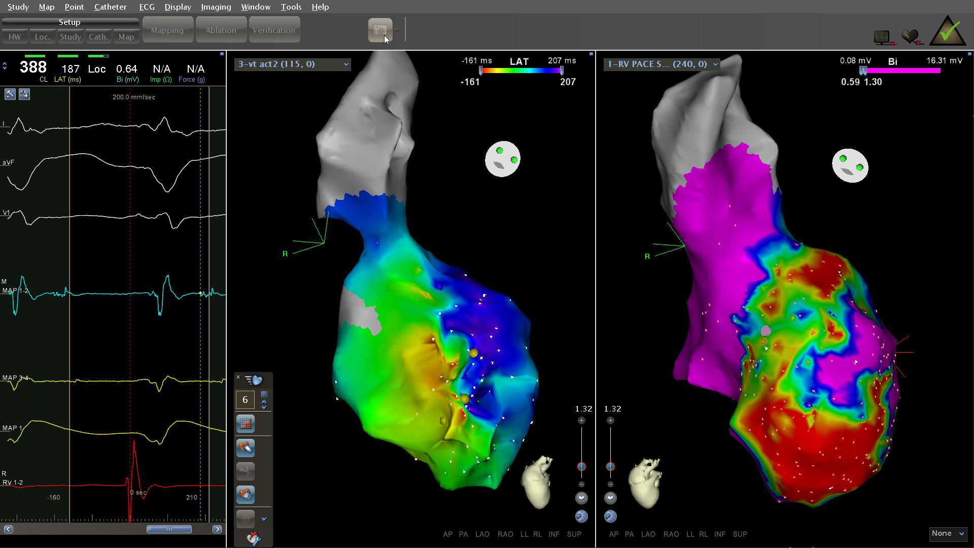
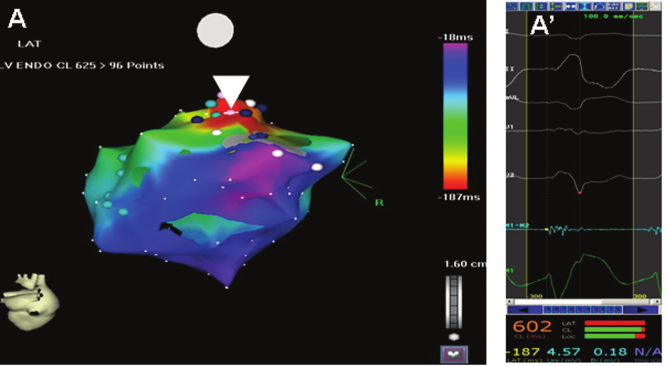
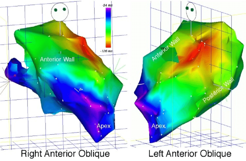
Activation map
- Isochronal map
- Activation time in relation to fixed reference
- Similar activation times marked with same colour
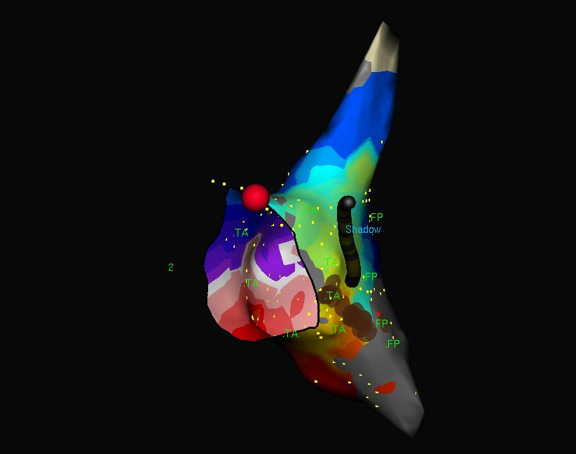
example - reentry
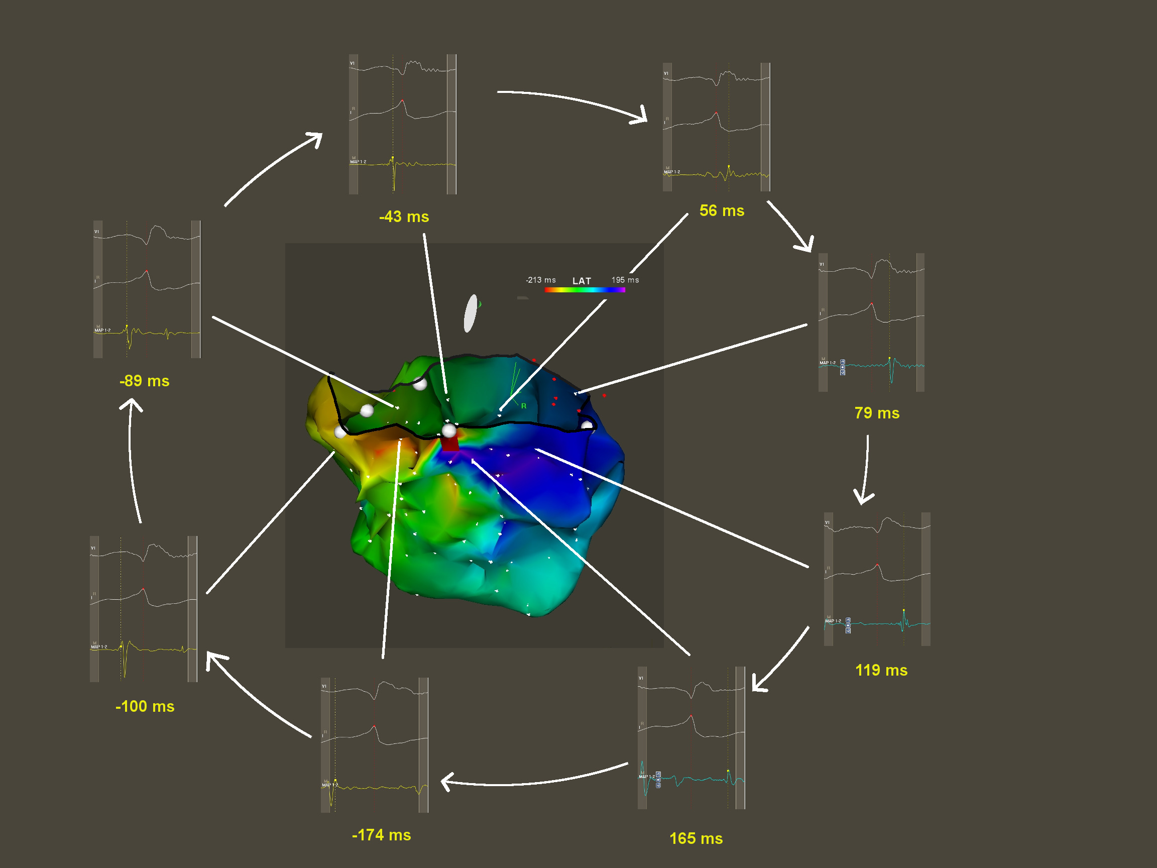
Kumar S, Subramanian A, Selvaraj RJ. Peritricuspid reentrant ventricular tachycardia in Ebstein's anomaly. Europace. 2014 Nov;16(11):1633
Assess conduction velocity
Combining information
Other maps
- Non contact mapping
- Manual tags
- Impedance maps
- Contact force
- Ripple maps
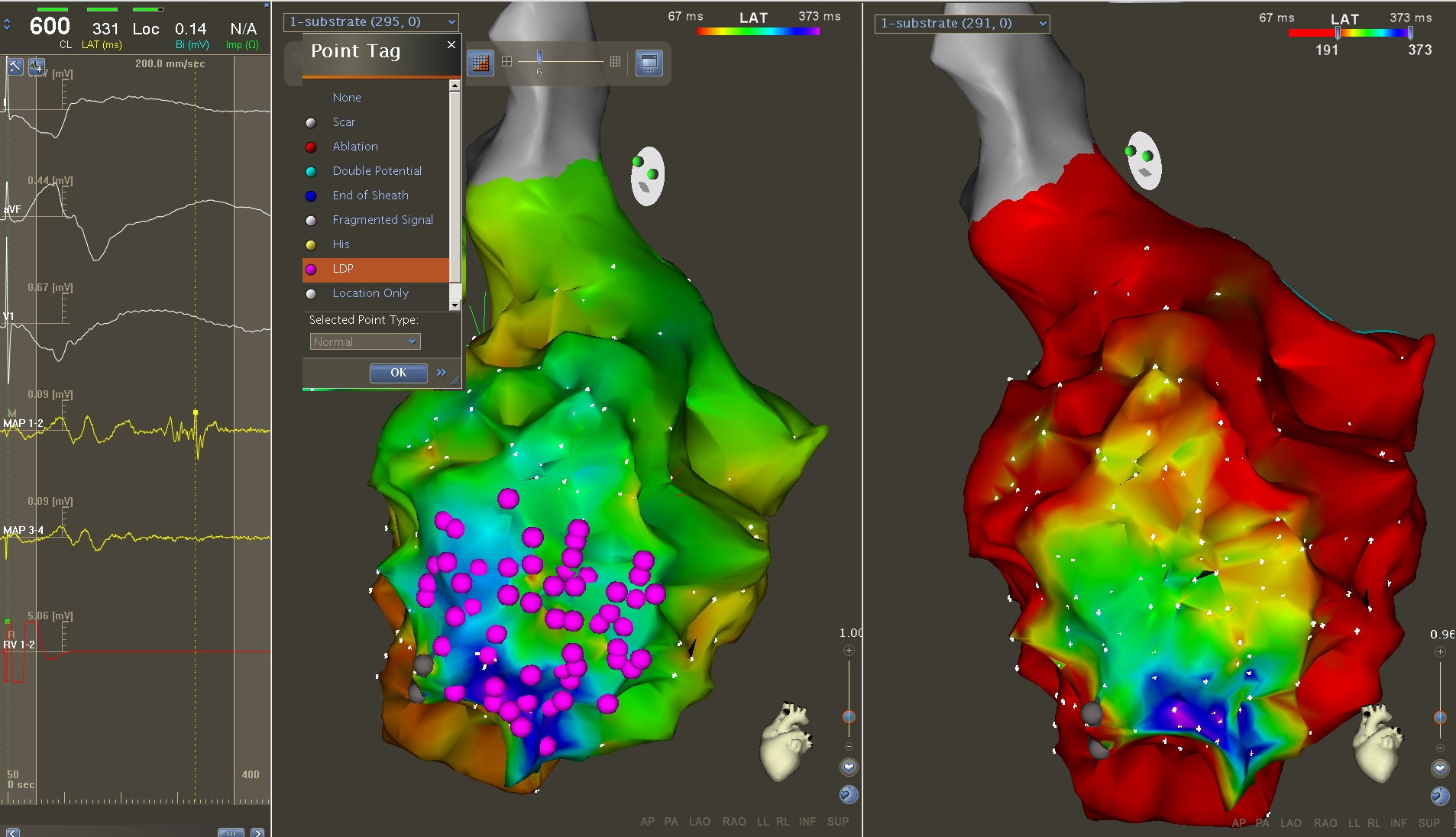
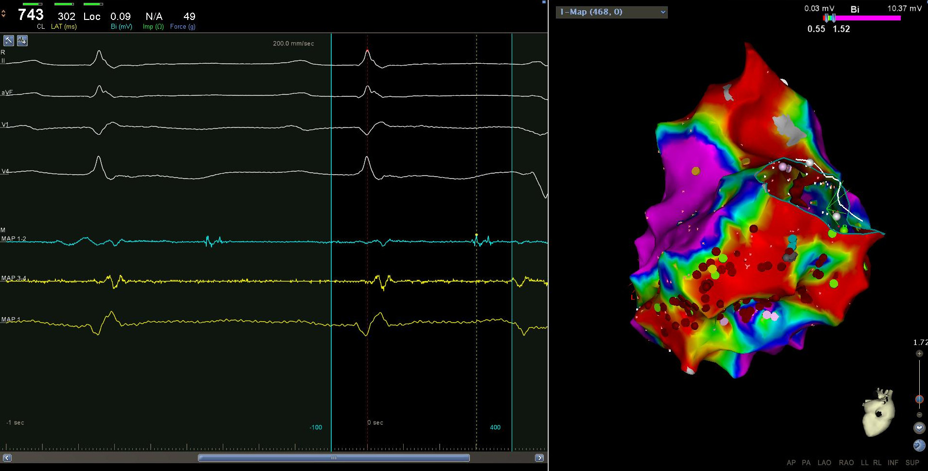
Substrate - Late potentials
Practical sequence
Choosing a reference
- Atrium
- fixed egm
- avoid multi-component signal
- screw in lead
- Ventricle
- Surface ECG QRS
- Intracardiac EGM
Setting the window
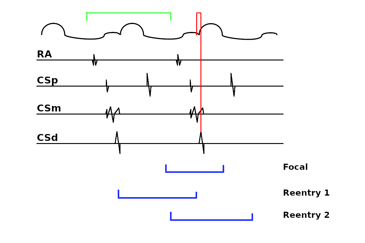
<n> focal = clear reentry = couple of choices
Window in reentry
- How big ? - 90% / 95% / 100%
- 10% of CL may be a large part of the circuit !
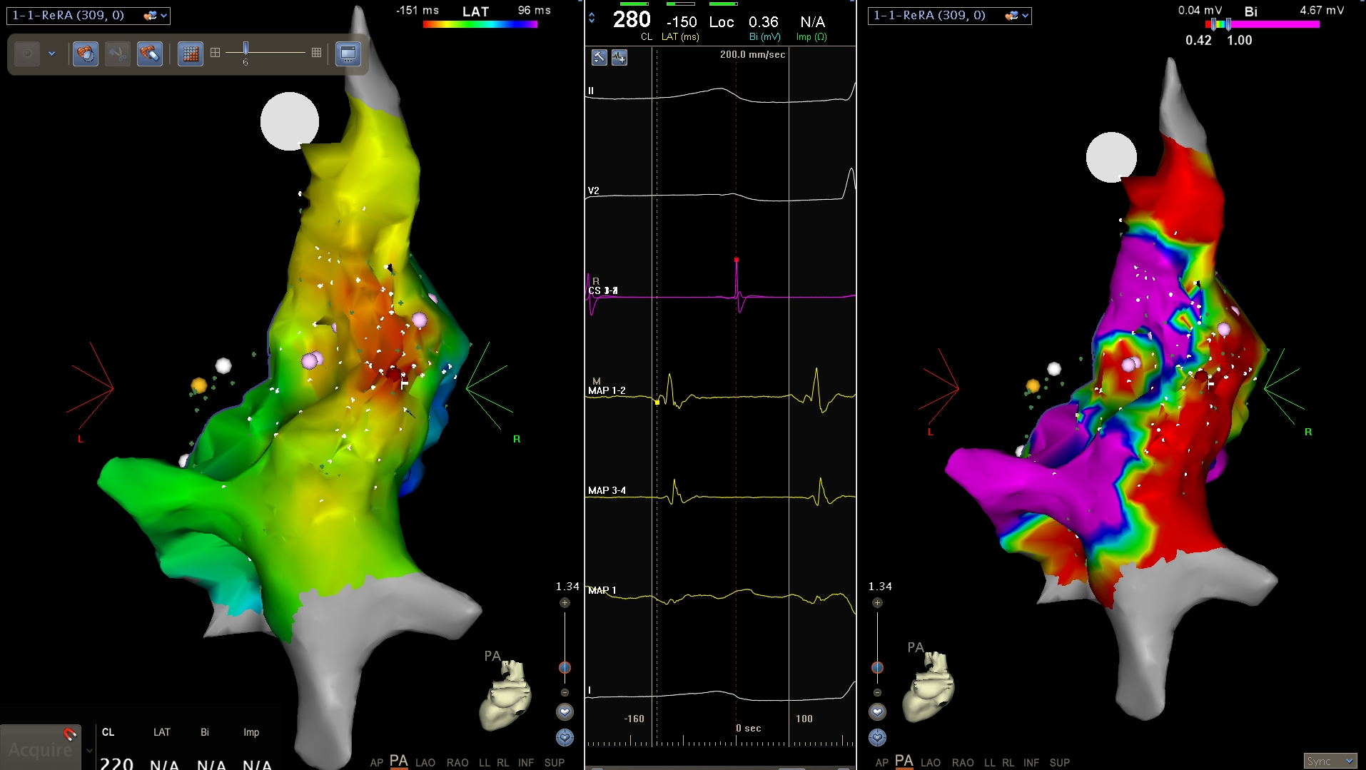
Two maps in trial flutter
Munoz F .. Asirvatham SJ. Three-dimensional mapping of cardiac arrhythmias: what do the colors really mean? Circ Arrhythm Electrophysiol. 2010 Dec;3(6):e6-11
Setting window in reentry
- Start in systole / diastole
- Window starting mid-diastole
- Early meets late at mid-diastole
- Identification of mid-diastolic activation
- De Ponti method
De Ponti R et al. Treatment of macro-re-entrant atrial tachycardia based on electroanatomic mapping: identification and ablation of the mid-diastolic isthmus. Europace. 2007 Jul;9(7):449-57
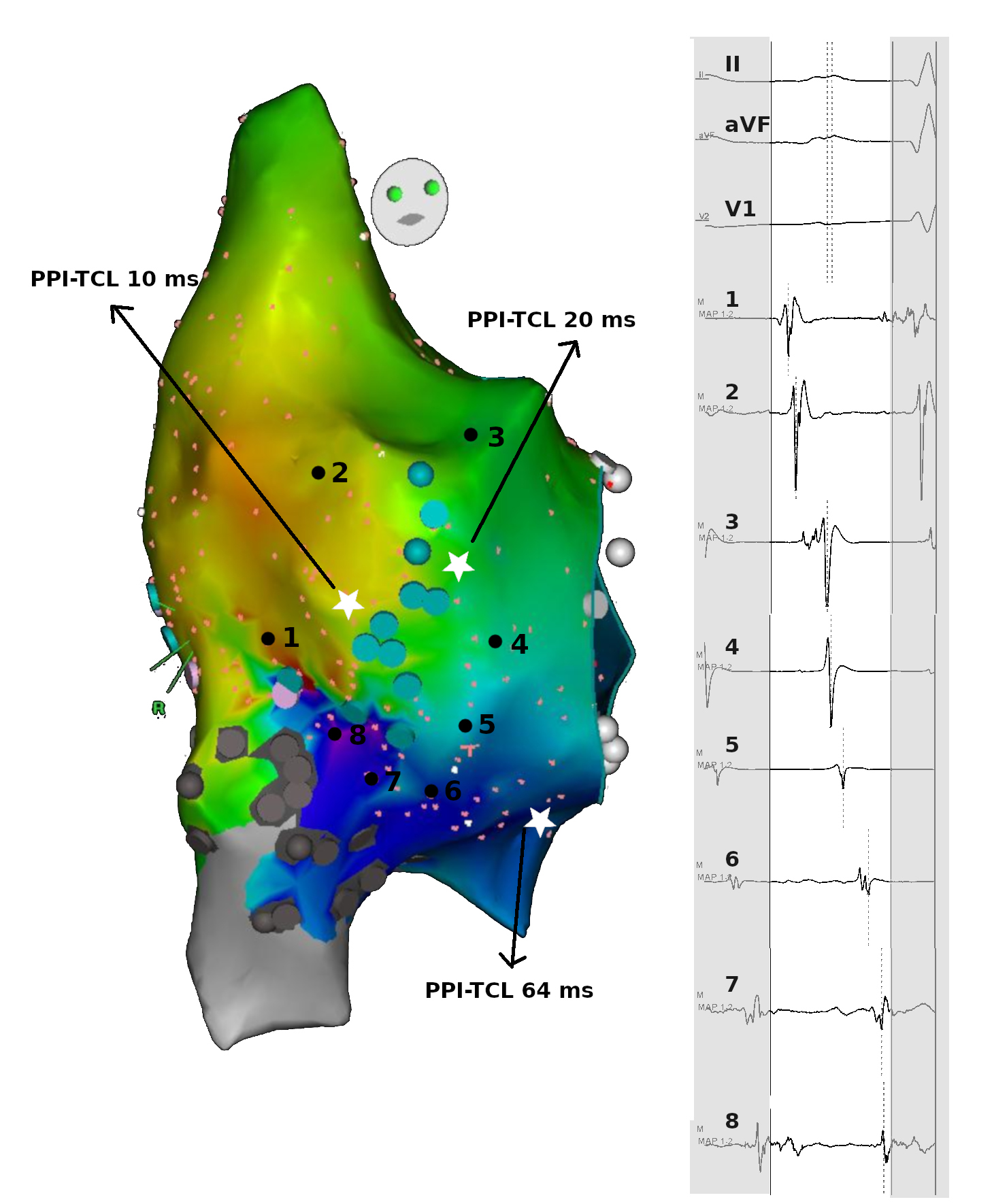
Identifying reentry
- Activation throughout the CL = LATs cover CL (example)
- Activation path as a circuit = Head meets tail
Setting window
Selvaraj RJ et al. Chasing red herrings: making sense of the colors while mapping. Circ Arrhythm Electrophysiol. 2014 Jun;7(3):553-6.
Setting window
Selvaraj RJ et al. Chasing red herrings: making sense of the colors while mapping. Circ Arrhythm Electrophysiol. 2014 Jun;7(3):553-6.
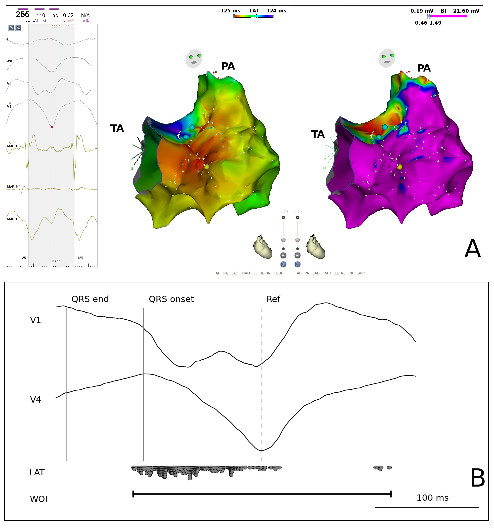
Setting window for substrate mapping
Manually collecting a point
- Catheter stability
- Contact
- See few beats
- Confirm beat taken
Beat buffer
Annotating activation
Annotating activation
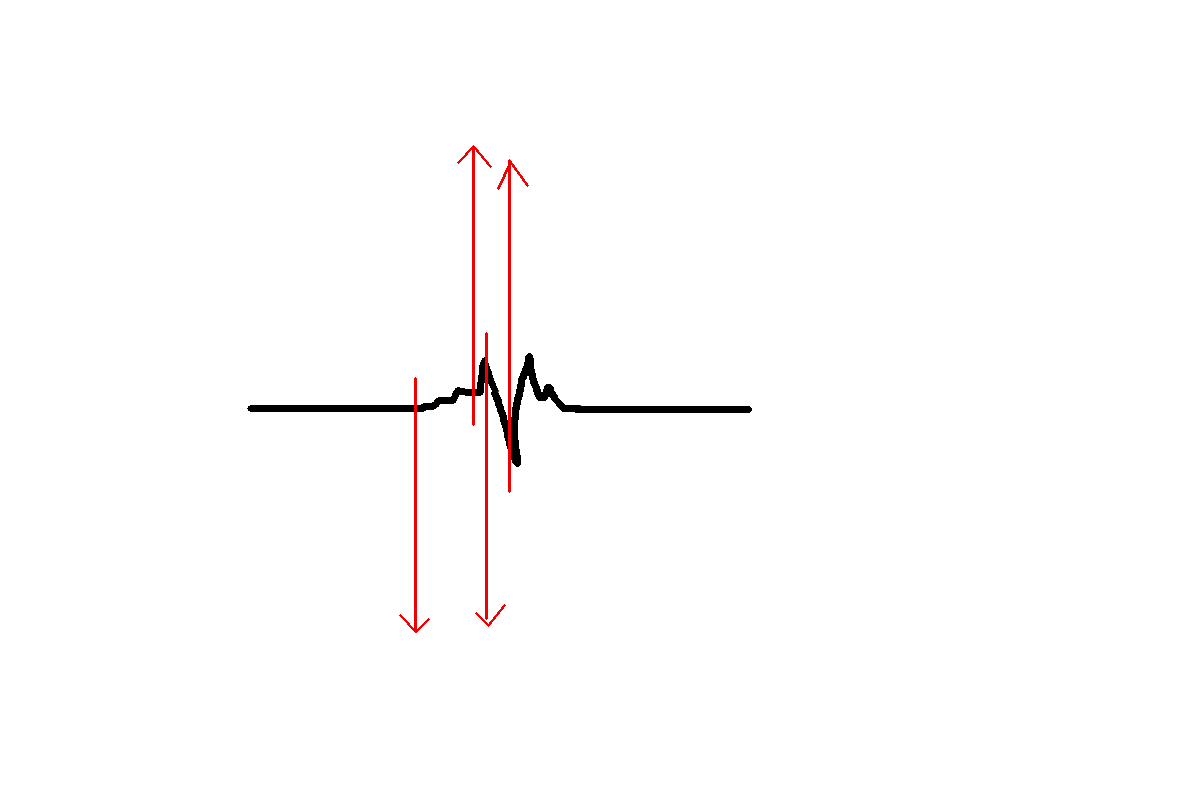
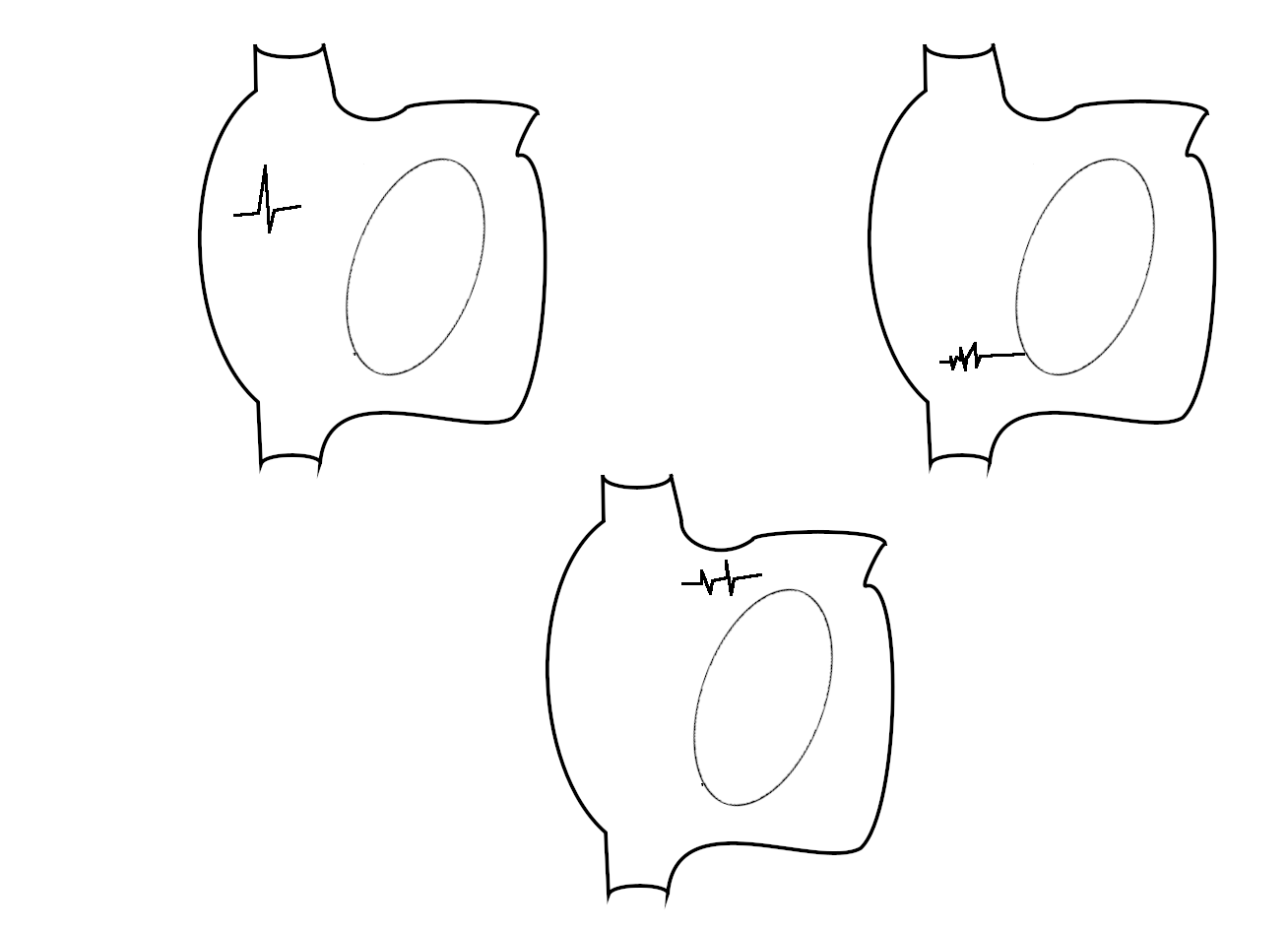
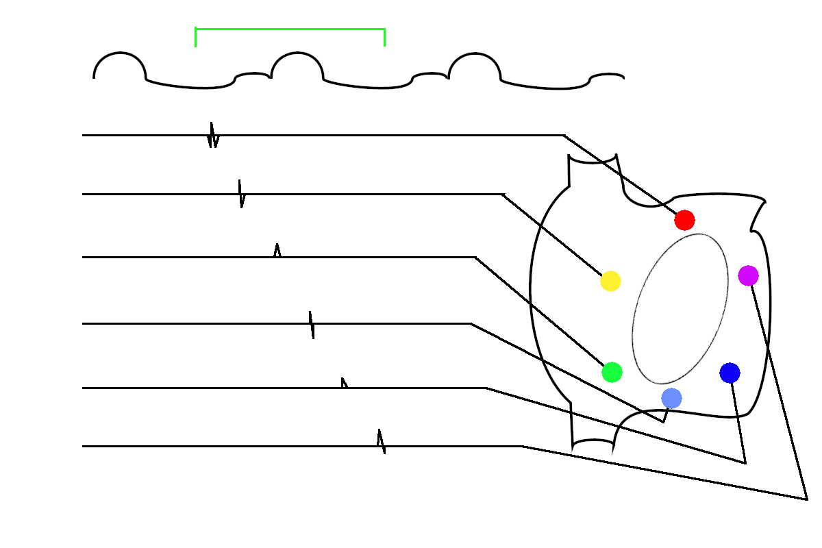
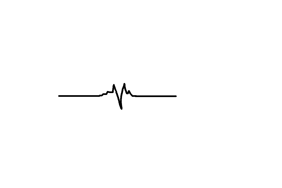
Annotating activation - single potentials (needs image)
- Earliest activation - may record far field
- Peak - May miss early activation
- Peak of first near field (Sam)
- Peak negative deflection (Nakagawa)
- Onset of first rapid deflection
- Have to correlate with neighbouring areas
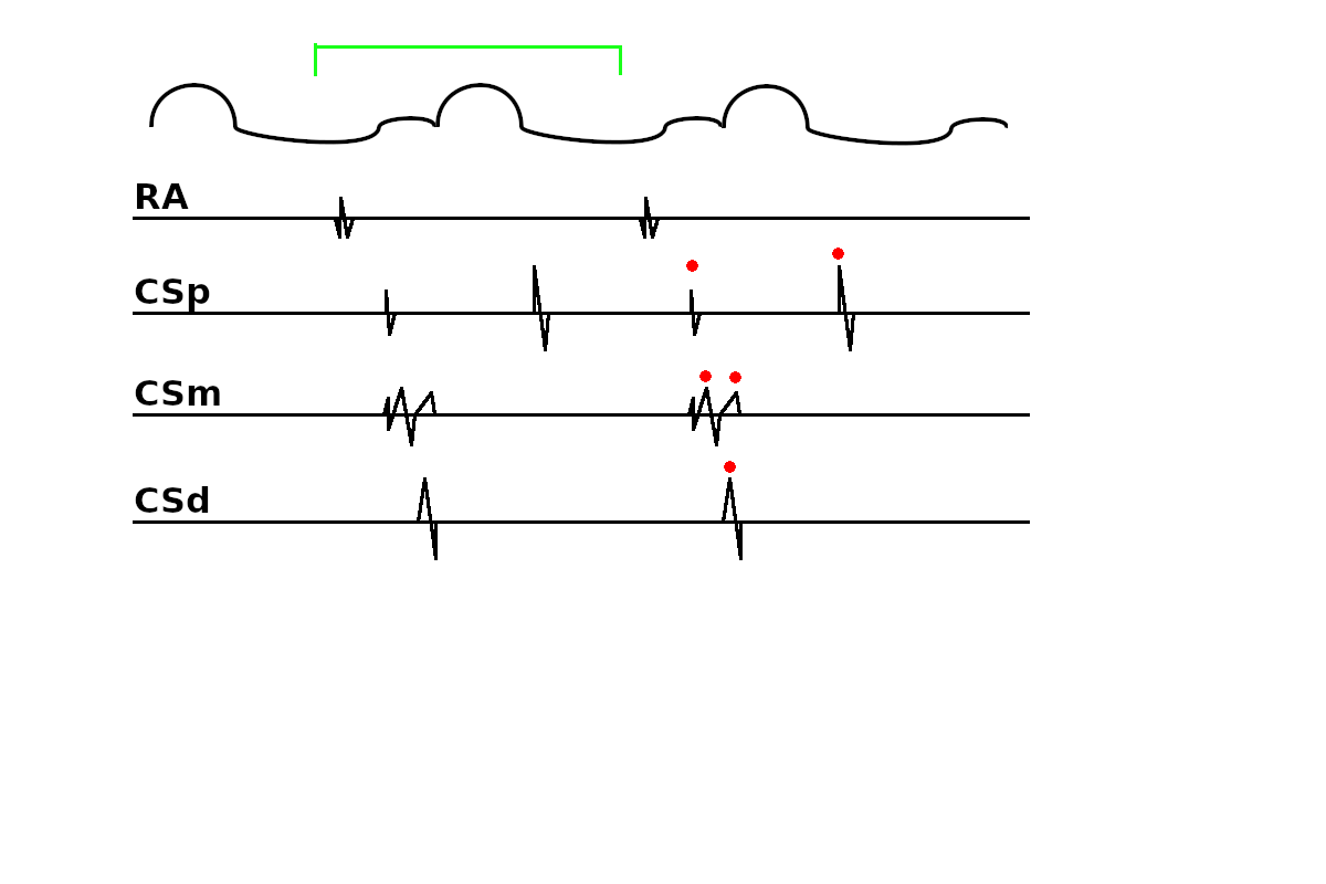
Annotating double potentials
- Attempt to identify the near field component
- Annotate consistently
- Tag as dp with location only when unsure or both appear near
Double potentials - near field
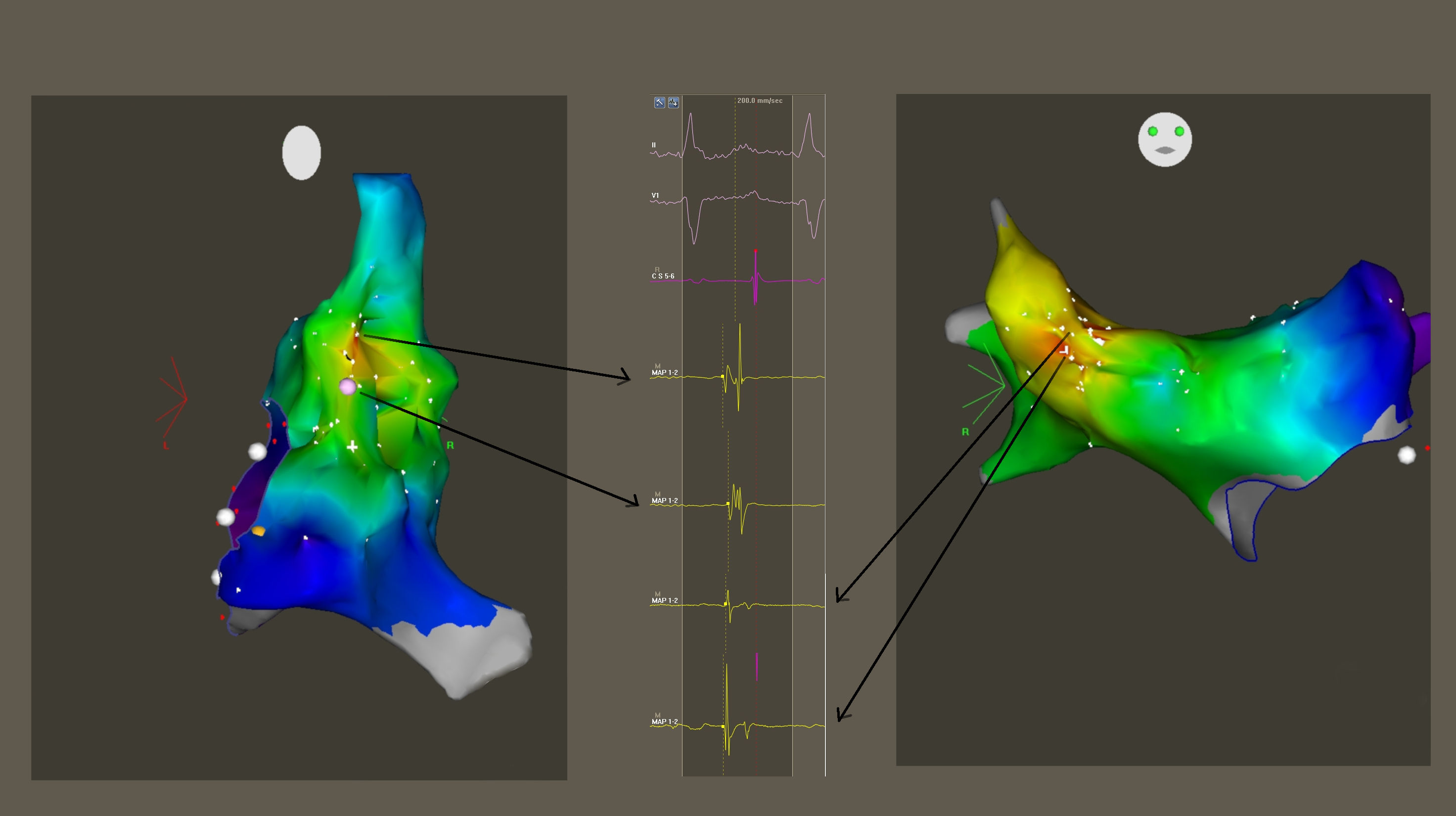
Selvaraj RJ et al. Which side are you on? - Deducing the chamber of origin of atrial tachycardia. Indian Pacing Electrophysiol J. 2017 Mar
- Apr;17(2):54-57.
Double potentials - near field
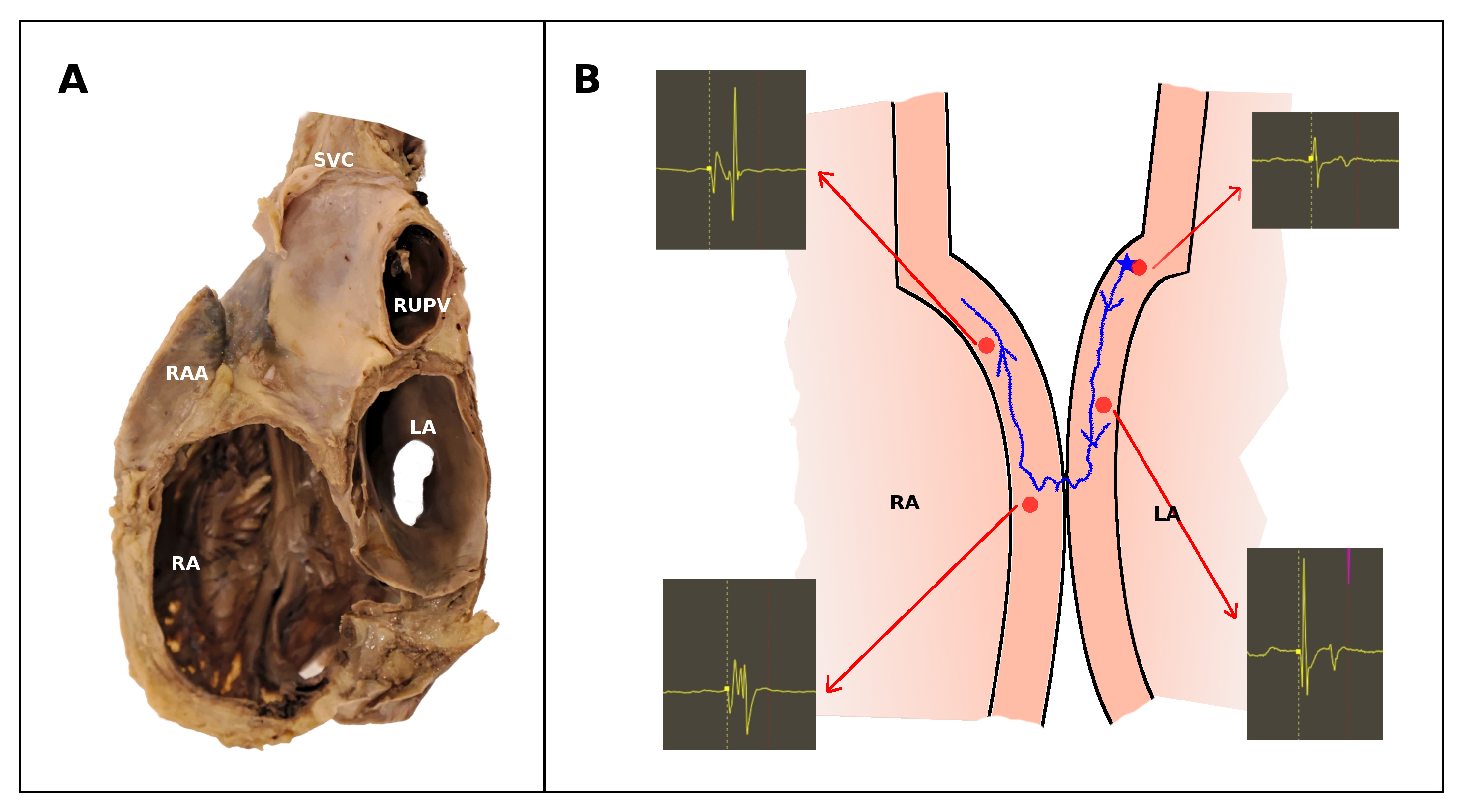
Selvaraj RJ et al. Which side are you on? - Deducing the chamber of origin of atrial tachycardia. Indian Pacing Electrophysiol J. 2017 Mar
- Apr;17(2):54-57.
Double potentials = pace to identify
Double potentials - tag
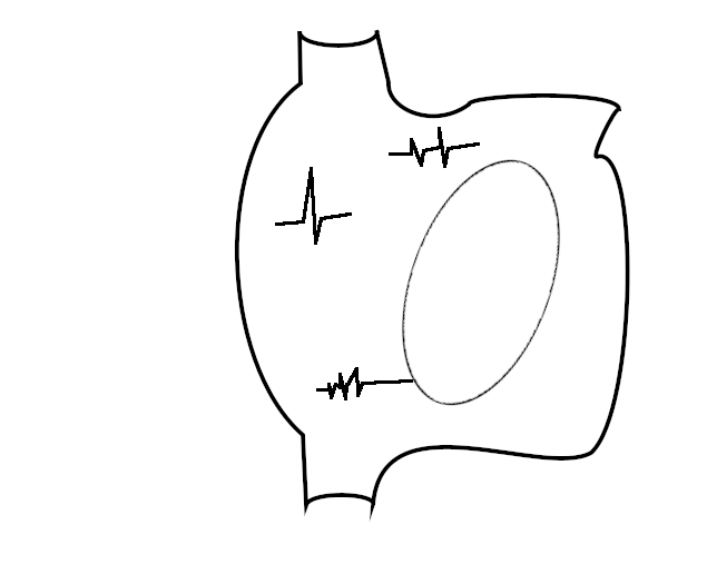
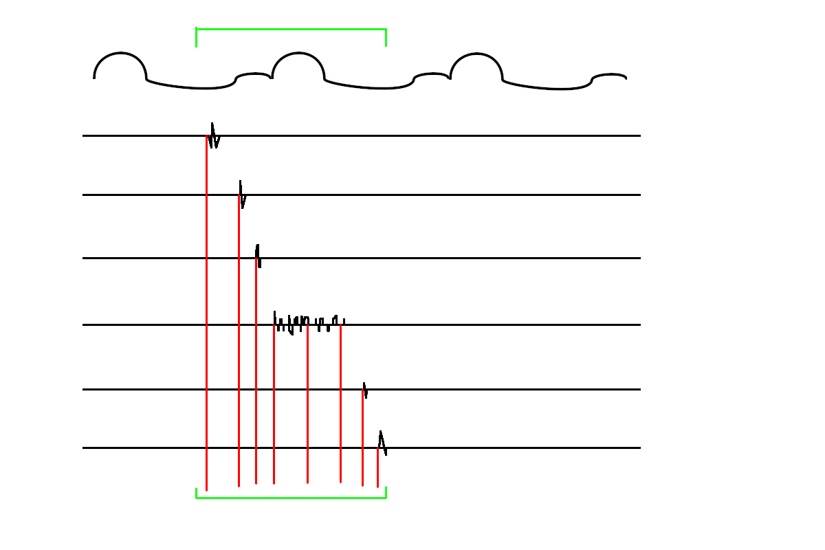
Annotating fragmented signals
- Consistent approach based on neighbours
- Adding single timing misleading
- Location only and add to CL later
- Multiple annotations with different times
Annotating fragmented signals
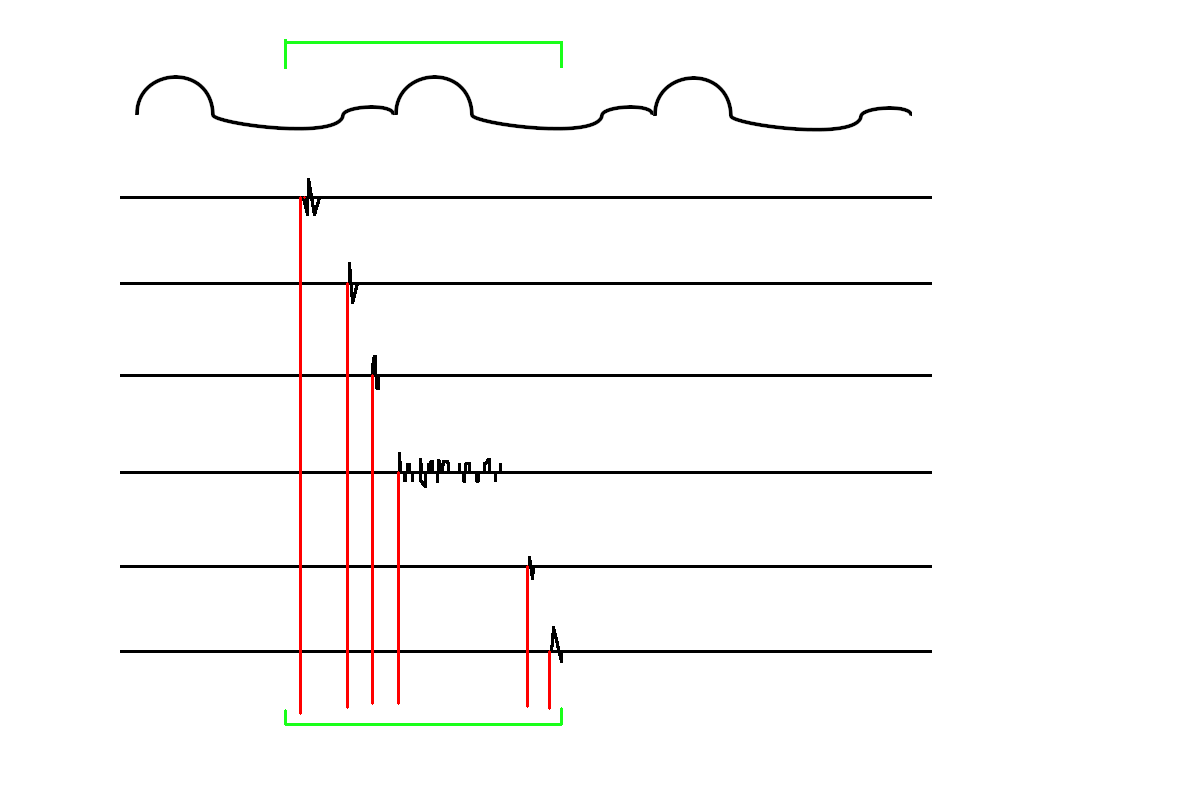
Annotating fragmented signals
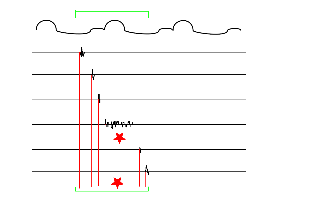
Annotating fragmented signals
Pitfalls
You only see what you have mapped
Samuel Asirvatham
You only see what you have mapped
Samuel Asirvatham
Interpolation
Summary
- Basics of 3D mapping
- Simplified some concepts
- Not covered recent advances