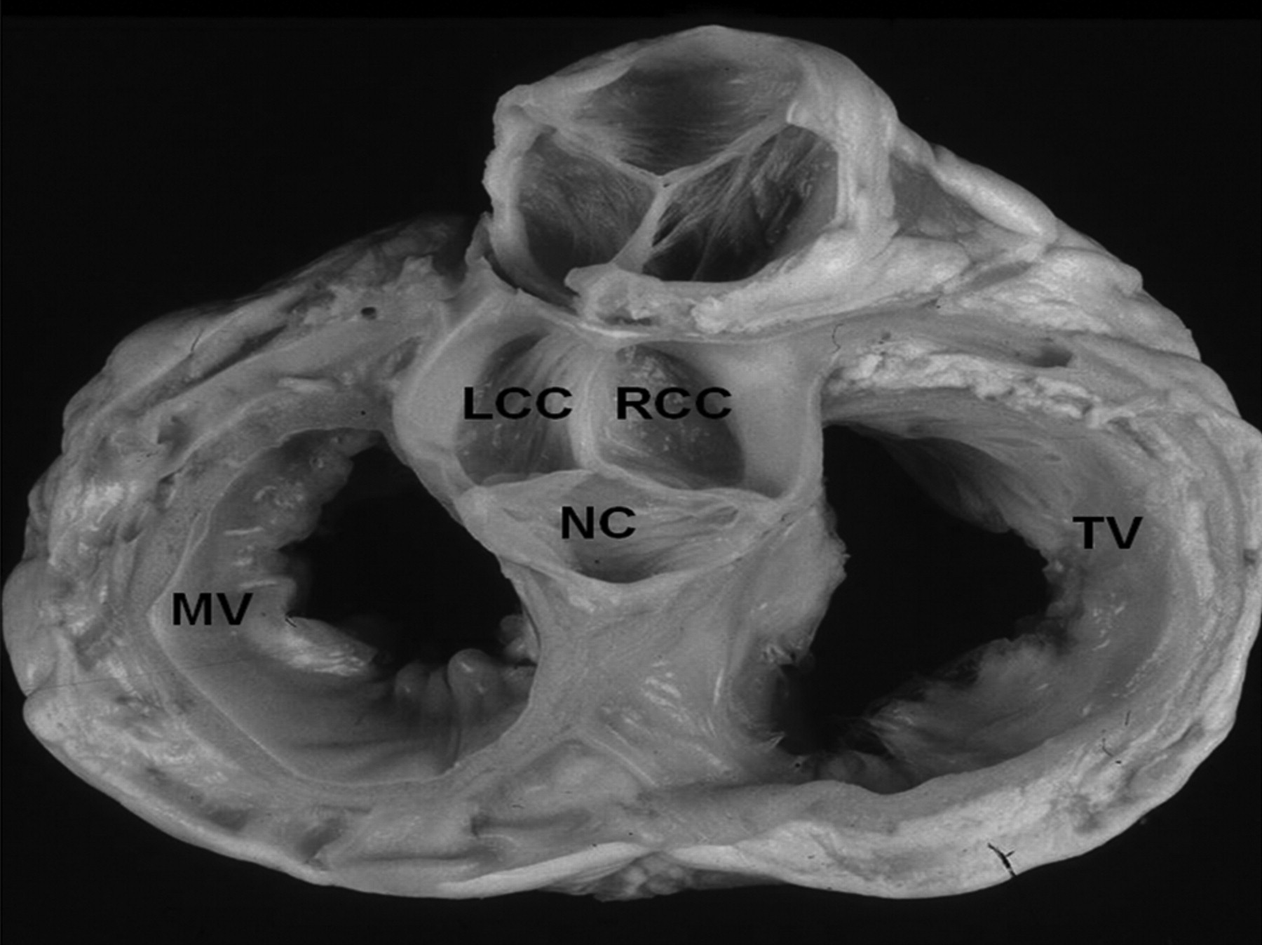
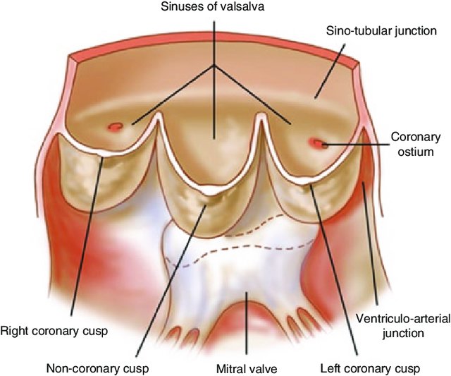
Anatomy
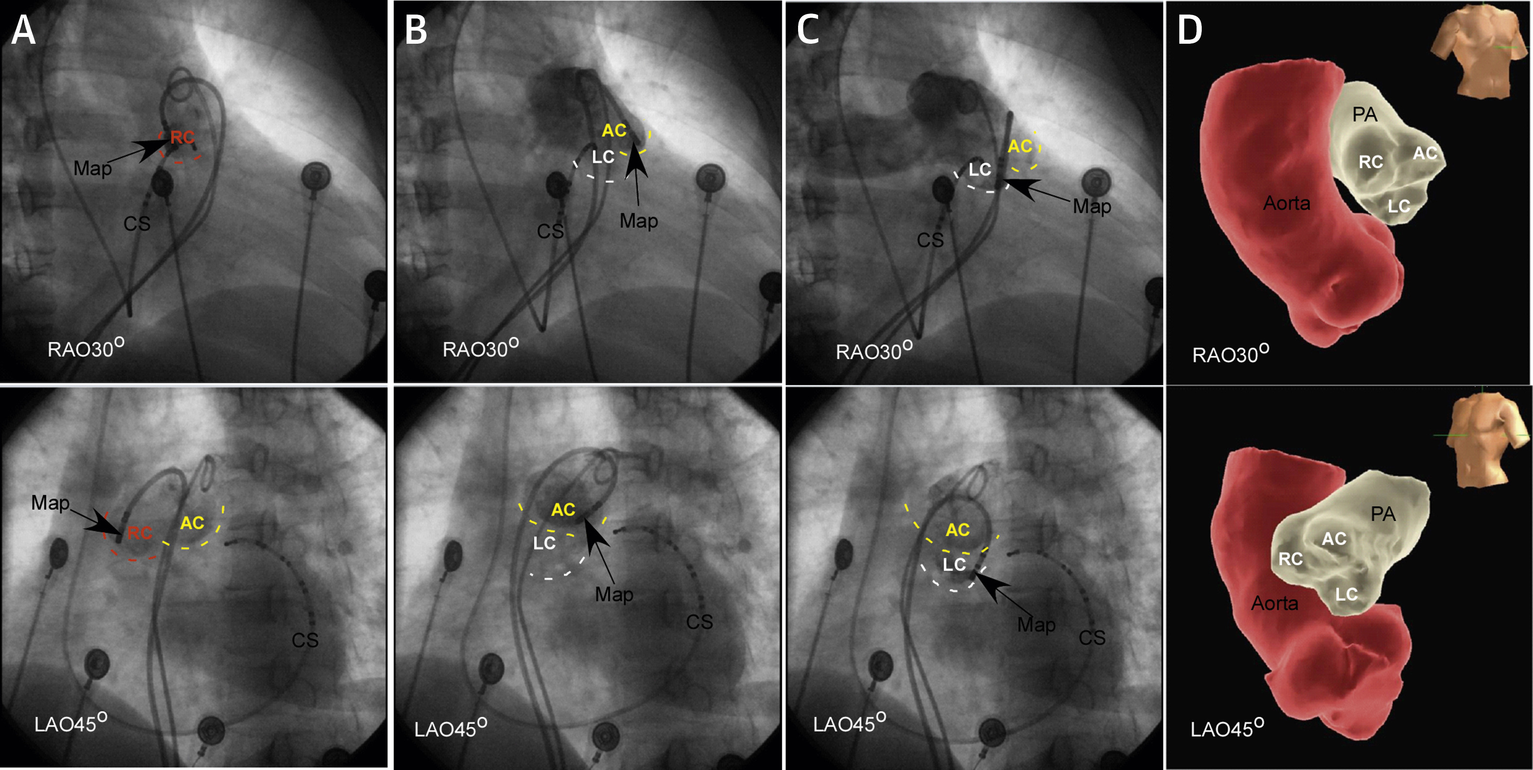
Outflow tracts relation
Niloufar Tabatabaei and Samuel J. Asirvatham. Circulation: Arrhythmia and Electrophysiology. 2009;2:316–326
LVOT
RVOT
Liao .. Ouyang. Idiopathic Ventricular Arrhythmias Originating From the Pulmonary Sinus Cusp: Prevalence, Electrocardiographic/Electrophysiological Characteristics, and Catheter Ablation. JACC 2015;66:2633-2644
RVOT
Vickram … Narasimhan. JOA 2020;36:471-477
ECG identification
Outflow tract
- Limb leads to identify OT origin
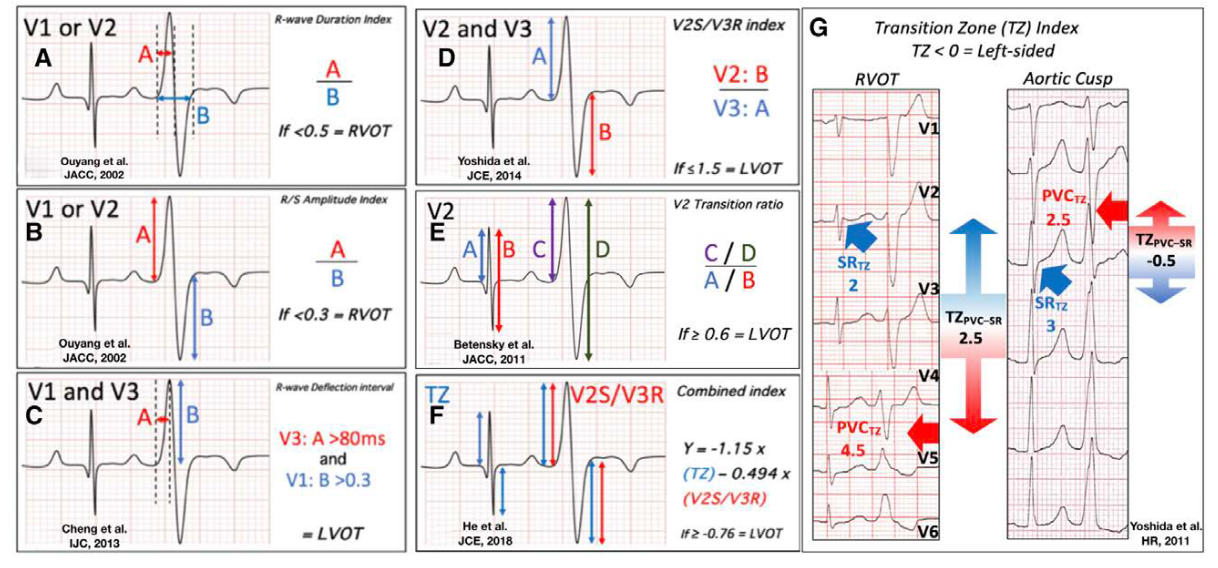
- Precordial leads to localize
V3 transition - RVOT or LVOT
Ouyang … Kuck. JACC 2002;39:500-508
ECG Algorithms
Anderson et al. Circ Arrhythmia 2019;12:e007392
PA-VA vs RVOT VA
- Higher R wave in inferior leads
- Higher aVL / aVR q wave ratio
- Increased R/S in V2
Y. Sekiguchi, K. Aonuma, A. Takahashi, et al. Electrocardiographic and electrophysiologic characteristics of ventricular tachycardia originating within the pulmonary artery J Am Coll Cardiol, 45 (2005), pp. 887-895
Coupling interval variability
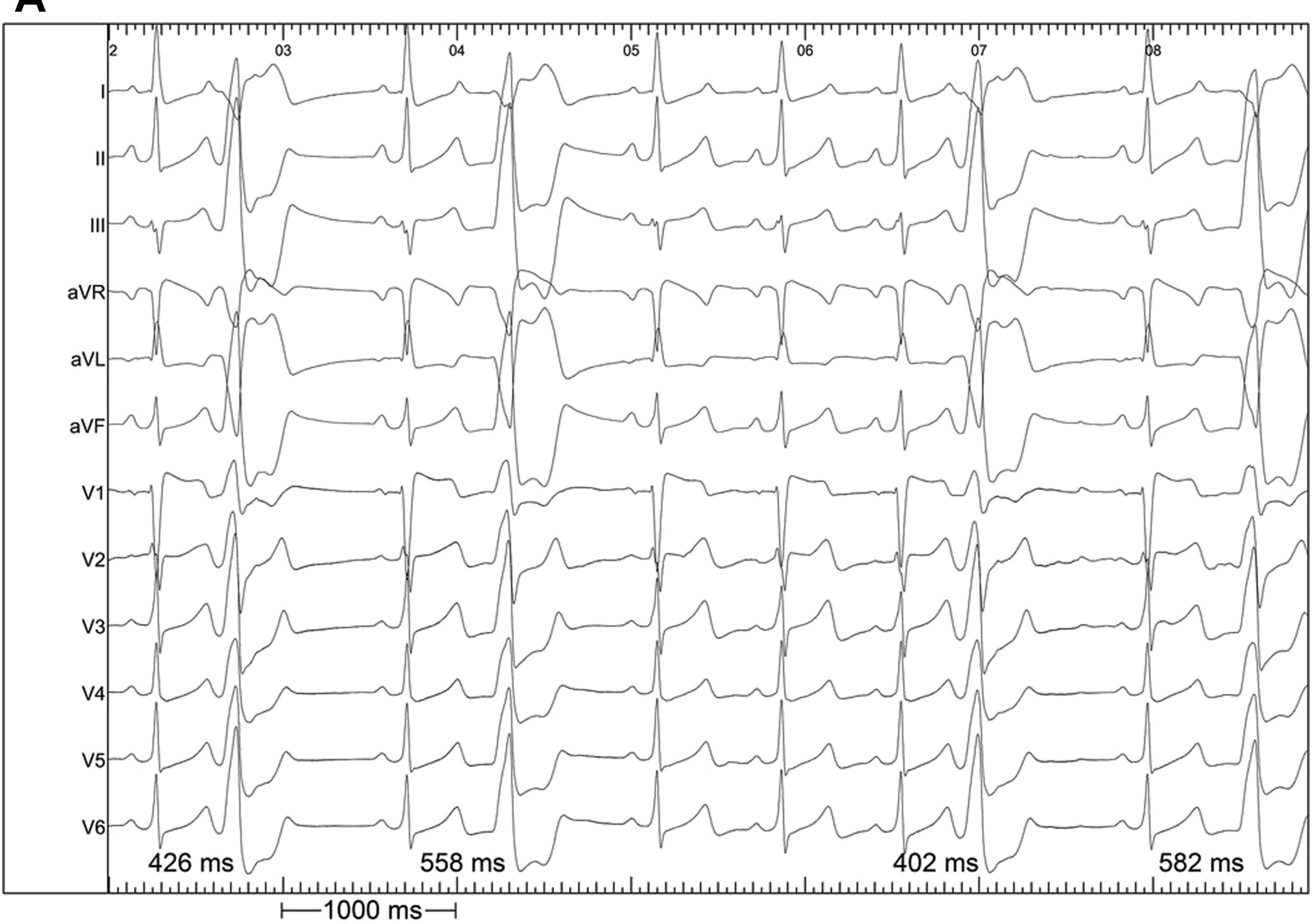
Bradfield JS, Homsi M, Shivkumar K, Miller JM. Coupling interval variability differentiates ventricular ectopic complexes arising in the aortic sinus of valsalva and great cardiac vein from other sources: mechanistic and arrhythmic risk implications. J Am Coll Cardiol. 2014;63(20):2151-2158. 10.1016/j.jacc.2014.02.551
EP identification
Start with a deeply placed CS catheter
Distinct EGM when mapping above the valve
- Muscle sleeve with fibrous / fatty tissue acting as substrate
- Typically seen above the aortic valve and pulmonary valve
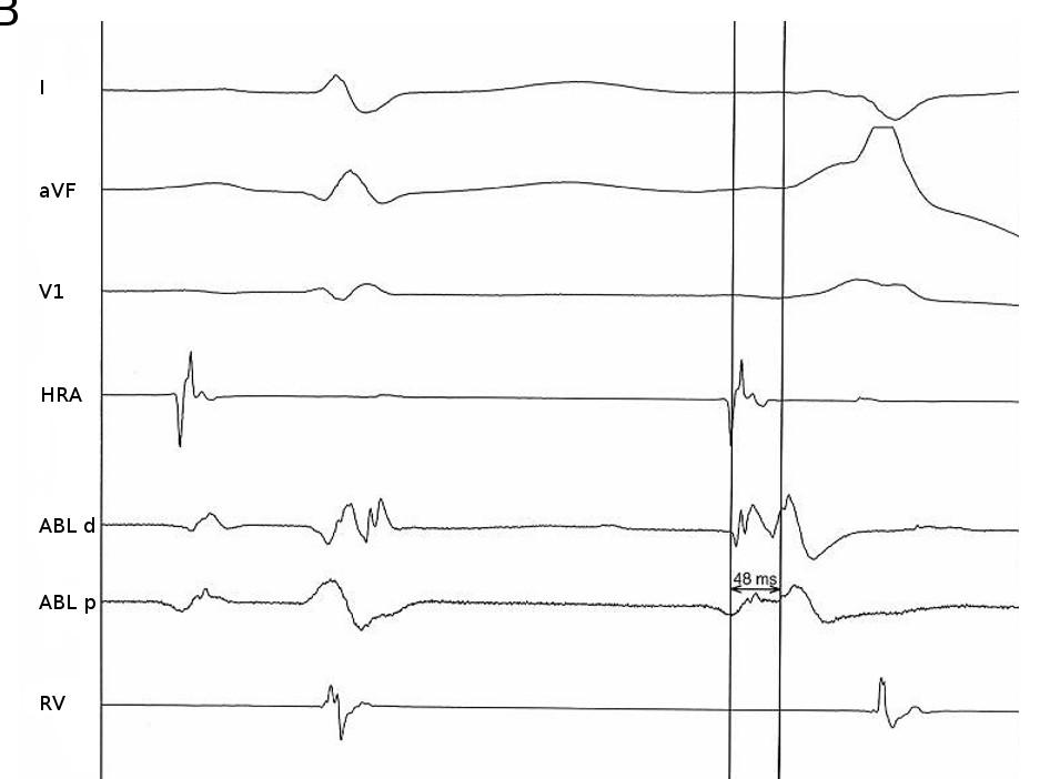
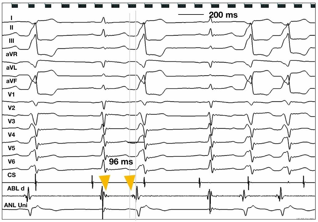
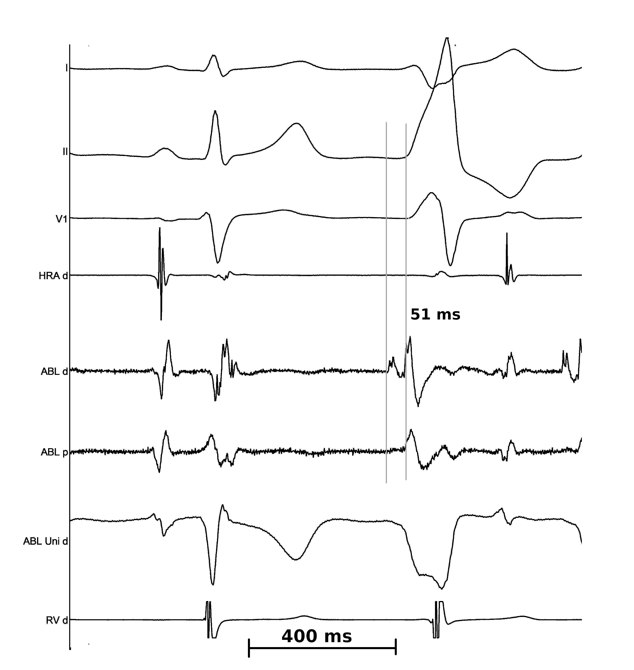
- Late in sinus rhythm and early before PVC
Komandoor S. Srivathsan et al. Mechanisms and Utility of Discrete Great Arterial Potentials in the Ablation of Outflow Tract Ventricular Arrhythmias. Circulation: Arrhythmia and Electrophysiology. 2008;1:30–38
Potential reversal in LCC
Selvaraj et al. Heart 2011
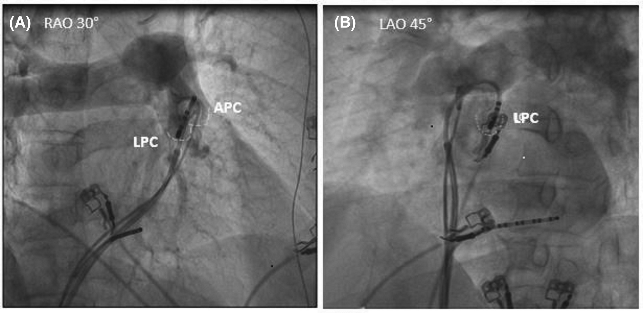
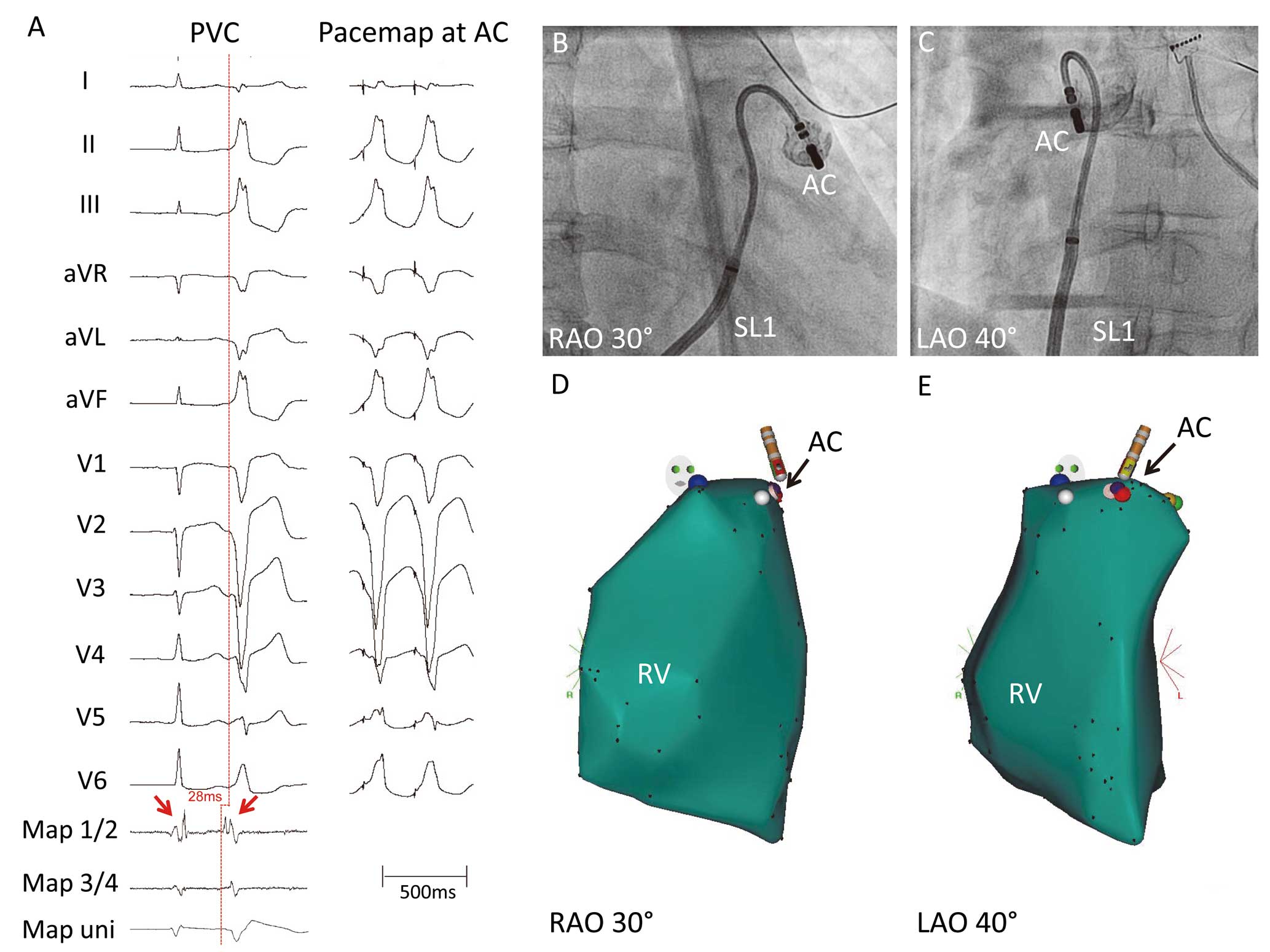
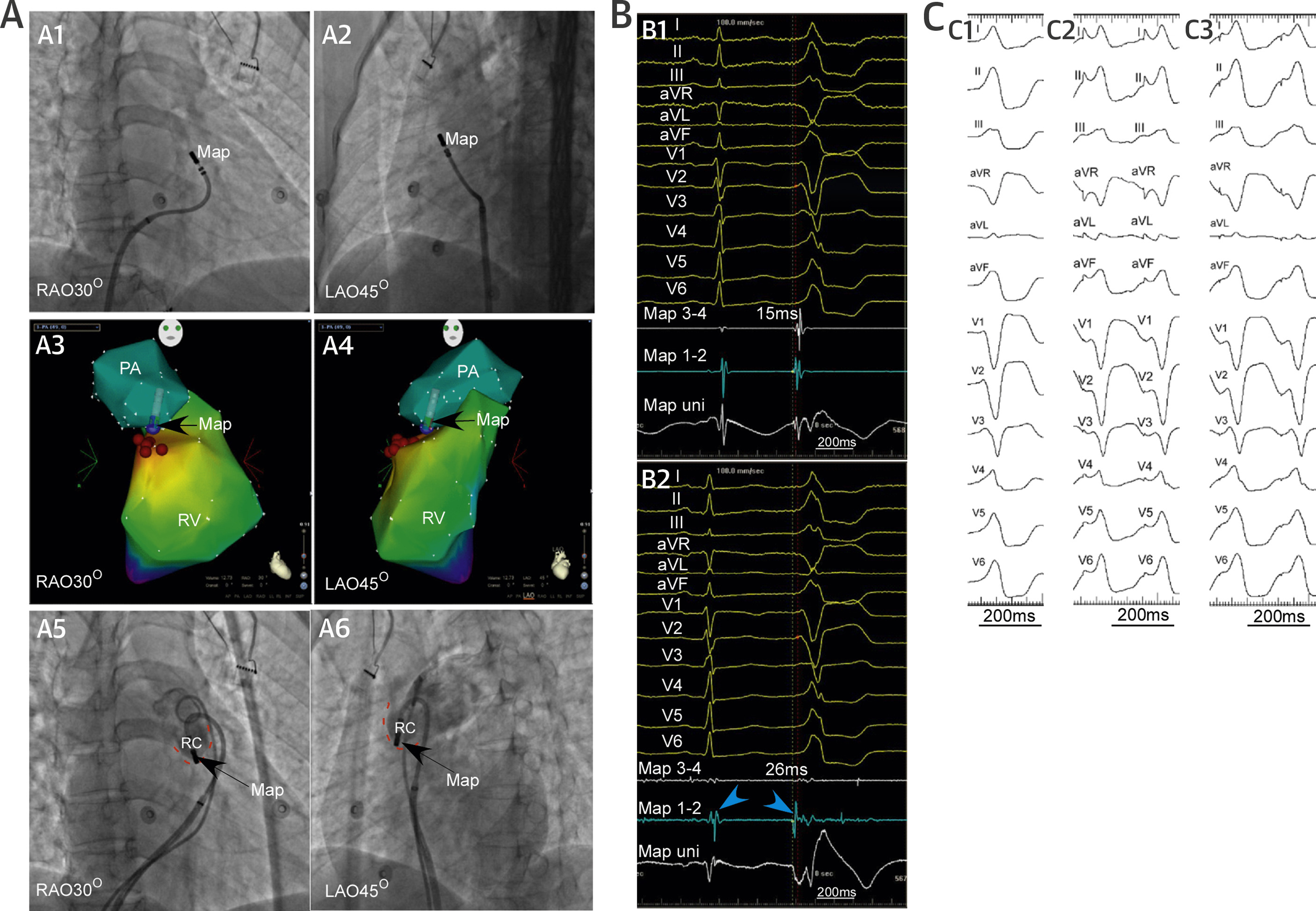
Pre potential in Pulmonary cusp
Vickram … Narasimhan. JOA 2020;36:471-477
Unipolar EGM
- Concordant with bipolar for RVOT without prior ablation
- May not concur in setting of prior ablation
- May not be useful while mapping above the valve
Conflict between pace mapping and activation mapping
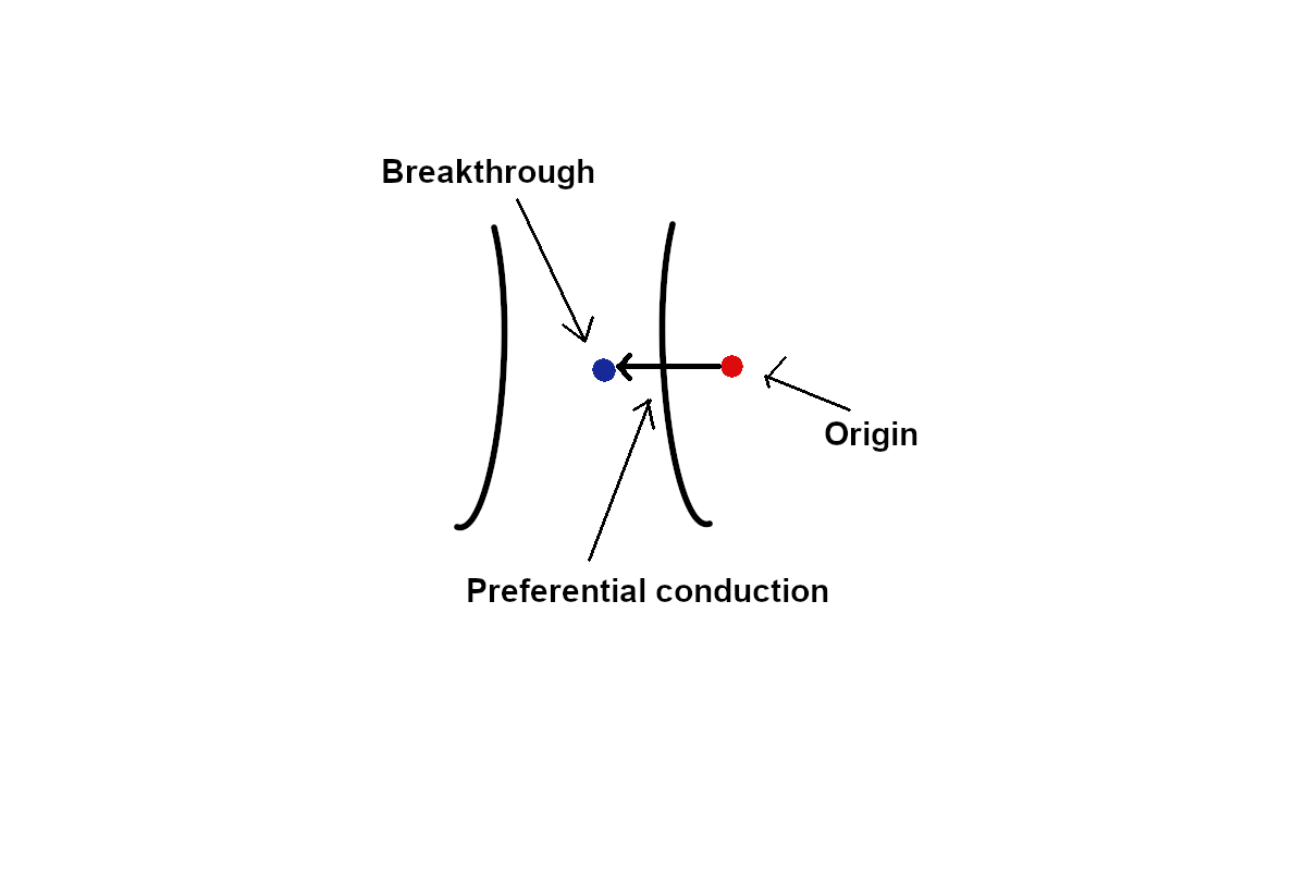
Wavefront spread
Csaba Herczku … Josep Brugada. Mapping Data Predictors of a Left Ventricular Outflow Tract Origin of Idiopathic Ventricular Tachycardia With V3 Transition and Septal Earliest Activation. Circulation: Arrhythmia and Electrophysiology. 2012;5:484–491
Ablation
Power
- 15-30 W may suffice
- Immediate cessation of PVCs indicator of success
Kabilan … Raja Selvaraj. Low power ablation for left coronary cusp ventricular tachycardia—Efficacy and long-term outcome. Indian Heart Journal 2018;17:S384-S388
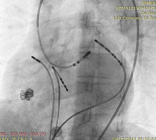
Ablation in coronary cusps - Aware of relationship to coronaries
Supravalvar RVOT VT
- Most from close to pulmonary valve
- Can be above the pulmonary valve level (1)
Timmermans C, Rodriguez LM, Crijns HJ, Moorman AF, Wellens HJ. Idiopathic left bundle-branch block-shaped ventricular tachycardia may originate above the pulmonary valve. Circulation 2003; 108: 1960–1967.
Most PA-VAs missed (thought as RVOT)
Liu … Lerman. Ubiquitous Myocardial Extensions Into the Pulmonary Artery Demonstrated by Integrated Intracardiac Echocardiography and Electroanatomic Mapping. Circ Arrhythmia 2014;7:691-700.
Pulmonary sinus cusp VT
Liao Z, Zhan X, Wu S, Xue Y, Fang X, Liao H, et al. Idiopathic ventricular arrhythmias originating from the pulmonary sinus cusp: Prevalence, electrocardiographic/electrophysiological characteristics, and catheter ablation. J Am Coll Cardiol 2015; 66: 2633–2644.
Suspect when unsuccessful in high RVOT
Summary
- Complex anatomy of the outflow tracts
- Differentiation between LVOT / RVOT
- ECG - Precordial transition, R in V1/V2
- EP - Activation in distal CS, signals in RVOT
- Awareness of OT VAs from above the valve