Introduction
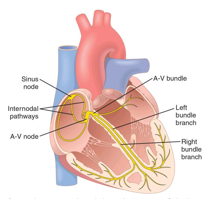
The heart is a two chambered organ
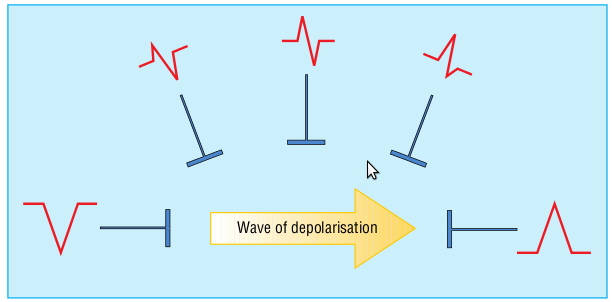
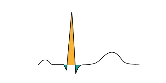
Anatomy of a wave
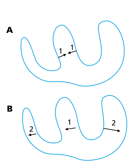
ECG deflection and wavefront
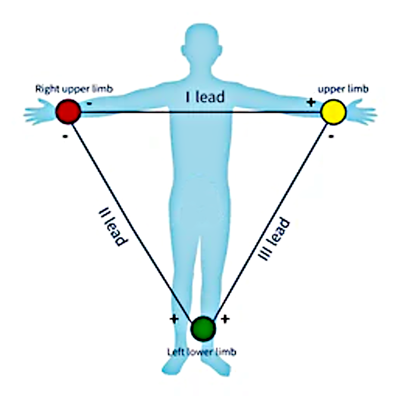
Limb leads
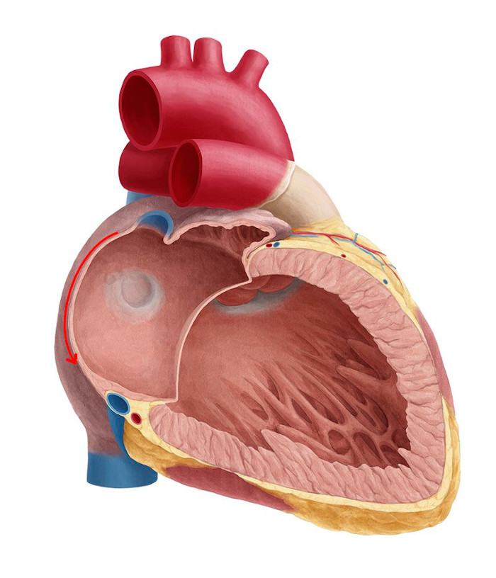
Wavefronts in atrium
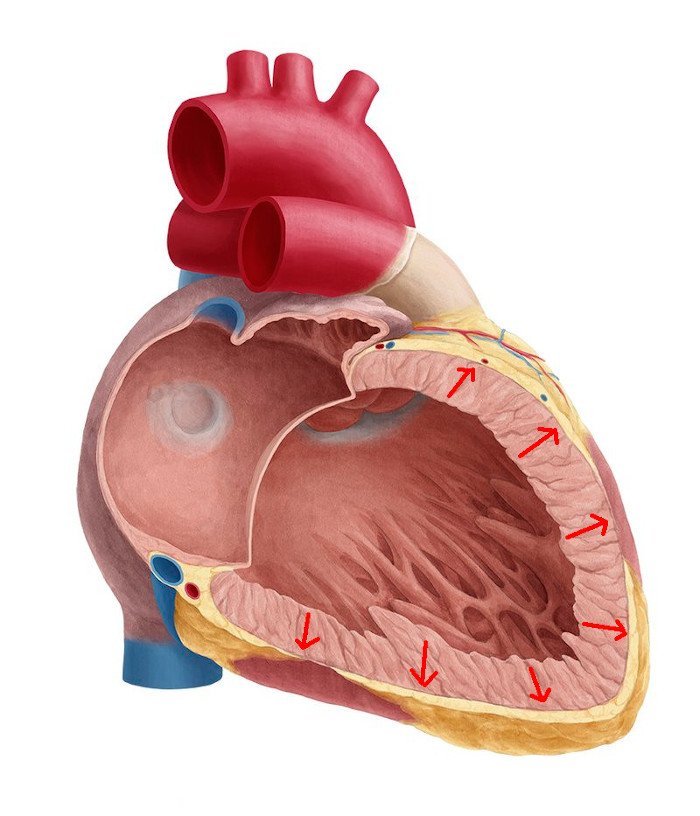
Wavefronts in ventricle
P wave
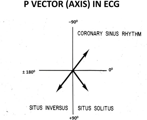
Atrial vectors
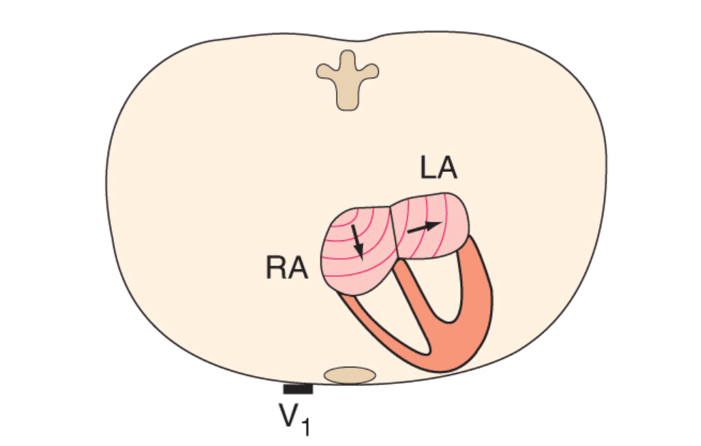
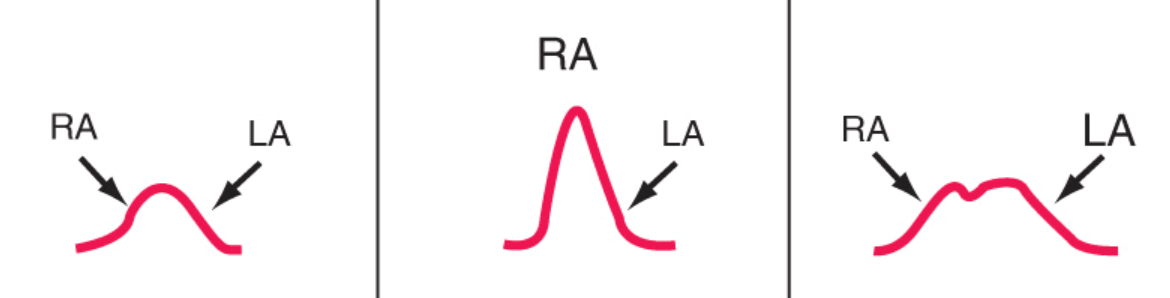
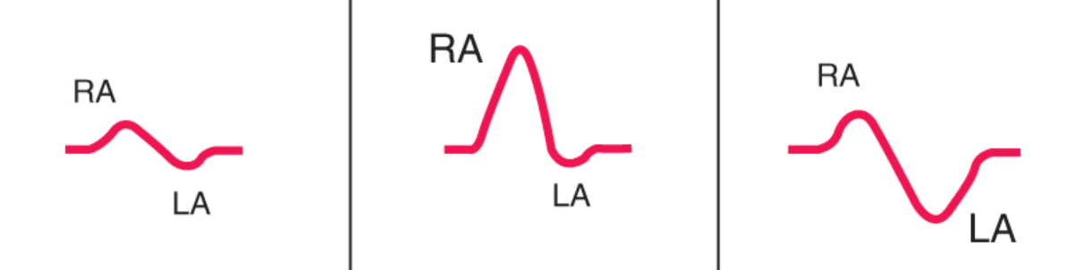
P wave - two atria
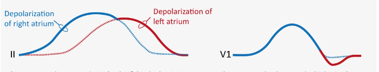
P wave lead II
P wave lead V1
Other P waves
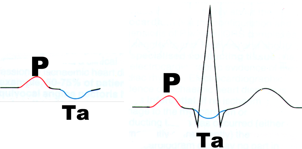
Ta wave
QRS and T waves
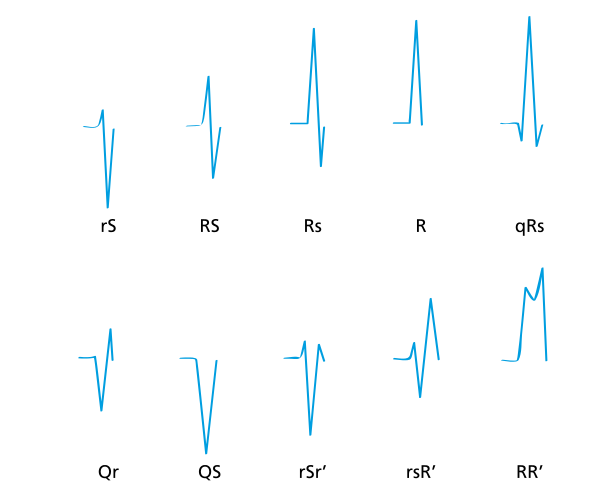
QRS - Terminology
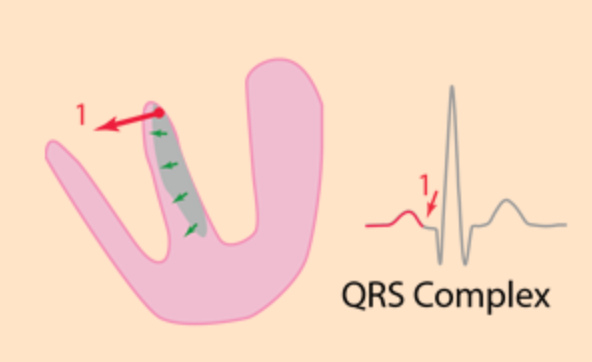
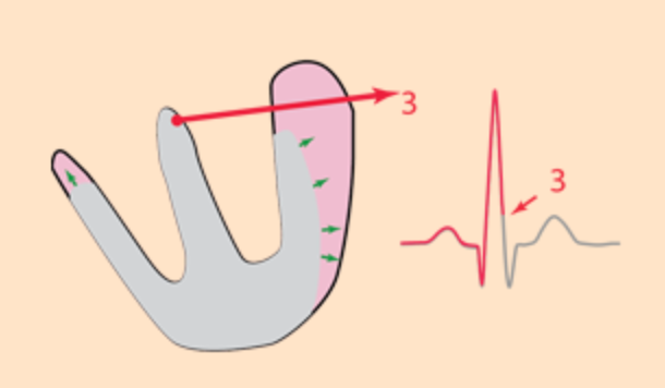
Ventricular activation
Septal q
Ventricular activation
Bundle branch blocks

T wave
Summary
What have we learnt ?
- Electrical activation occurs as waves of depoalrisation and repolarisation
- Wavefronts moving towards or away from recording electrode produce a deflection in the ECG
- Deflections produced by the atrium and ventricle can be used to infer the activation patterns
- Forms the basis for all further discussion on the ECG