![]()
Learning Objectives
- Understand basics of pacemaker function
- ECG evaluation of pacemaker function
- Interpretation of paced ECGs - When to worry / refer
General principles of pacemaker function
Pacing
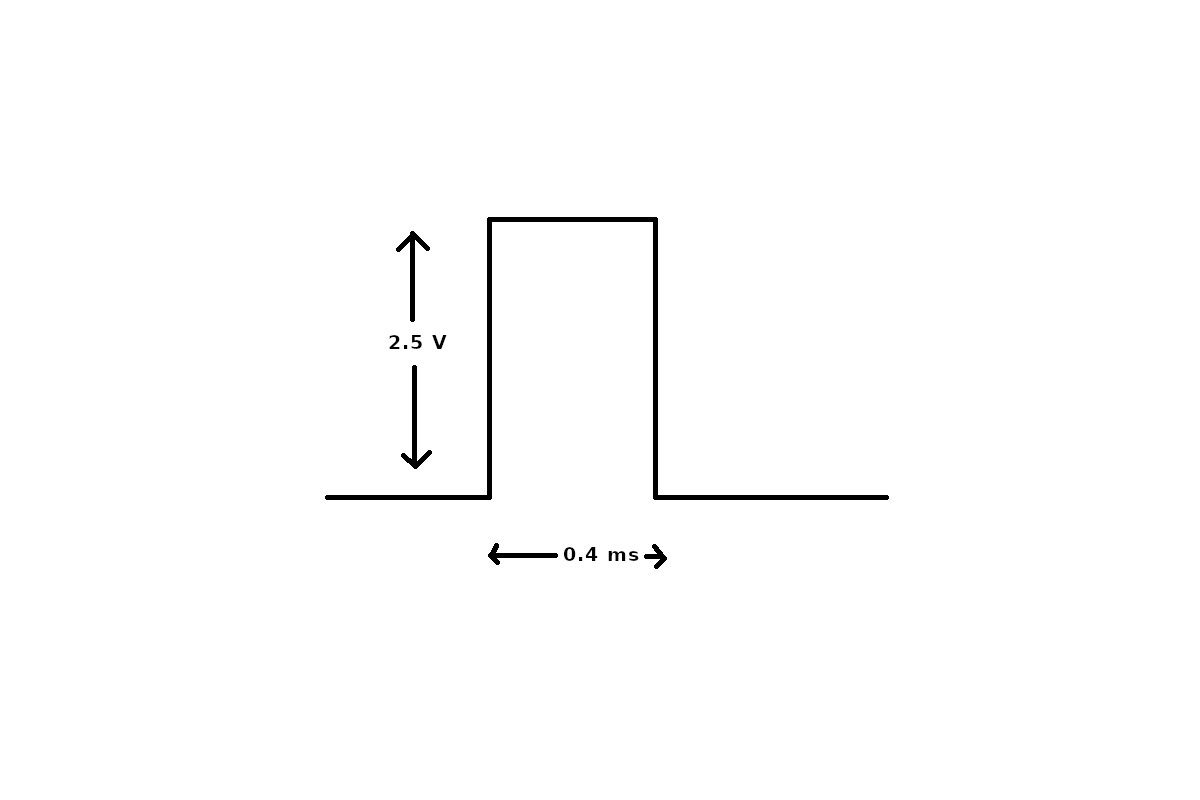
- Delivers a rectangular electrical stimulus at timed intervals
- Seen as a high frequency pacing spike
- Followed by capture of the respective chamber
Pacemaker pulse
Pacing
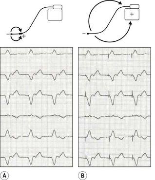
Unipolar / bipolar pacing
Pacing
Asynchronous pacing - VOO
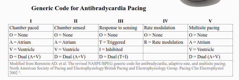
The pacemaker codes
Sensing
- Watches for intrinsic activity
- Generally inhibits pacing (resets timer)
- For dual chamber pacemaker, sensing in atrium initiates pacing in ventricle after delay
Sensing
Sensing - VVI
Event markers
Timing
A Systematic Approach to Pacemaker ECG
Step by step
- What is the rate and rhythm ?
- What is the underlying rhythm (sinus rate / AV conduction) ?
- What pacemaker ?
- Pacing function at each location ?
- Sensing function at each location ?
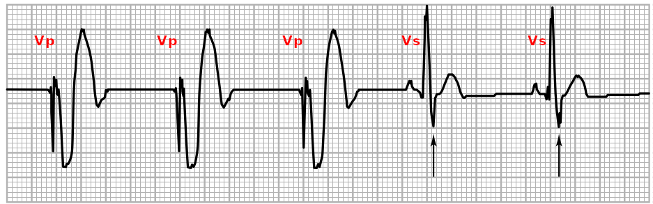
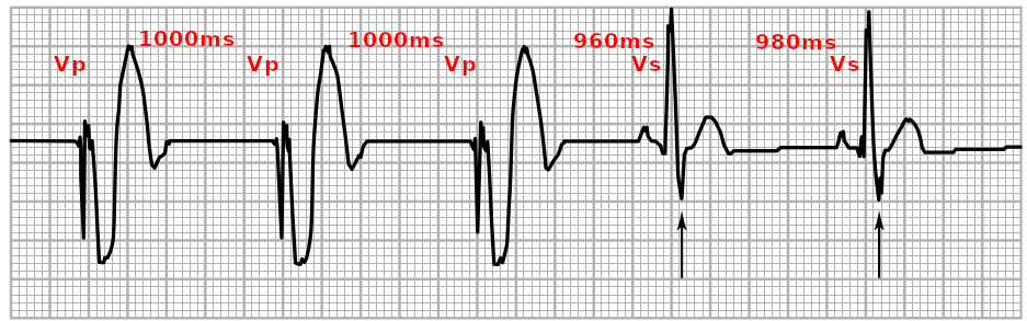
Pacing function
- Pacing - Delivery of pacing spike on time
- Capture - Myocardial capture by pacing spike
Sensing function
- Correctly identify only true intrinsic activity
- Oversensing - Not pacing when it should
- Undersensing - Pacing when it should not
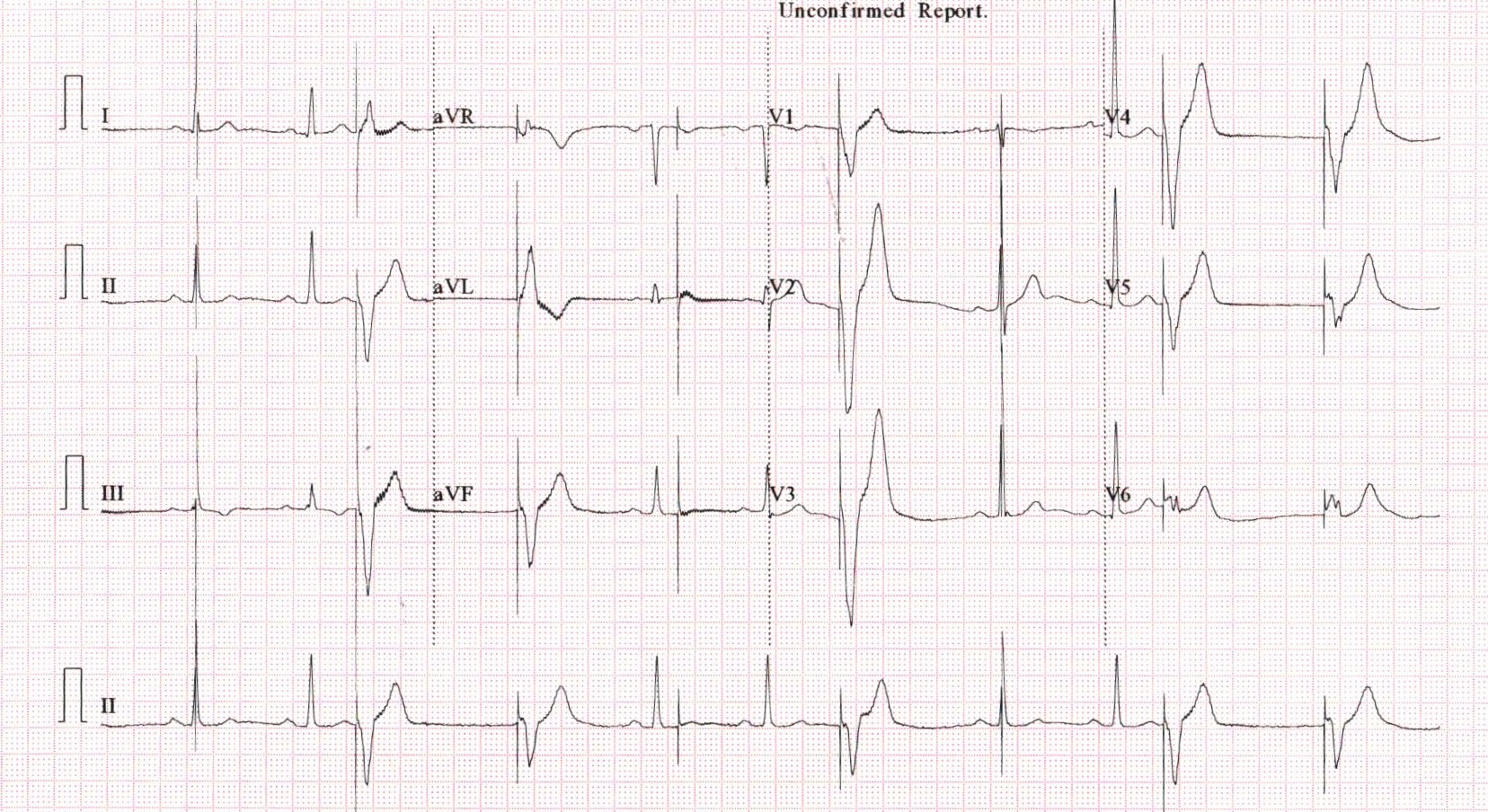
Lets look at some ECGs
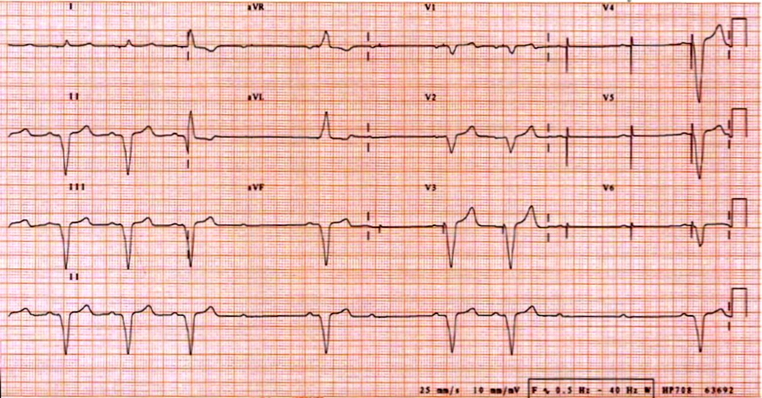
Pacemaker implanted 2 years back - Referred as "pacemaker not working"
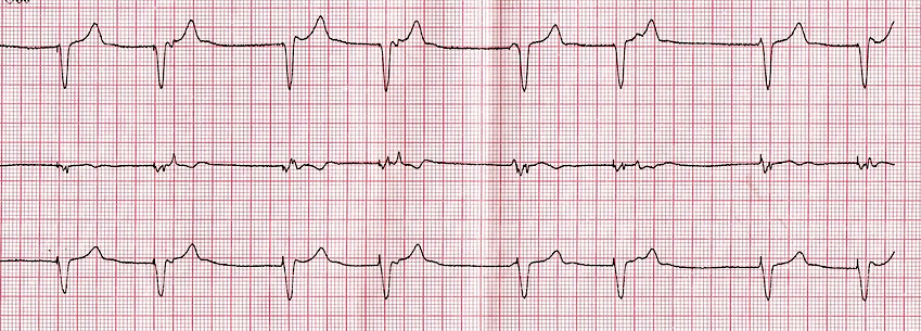
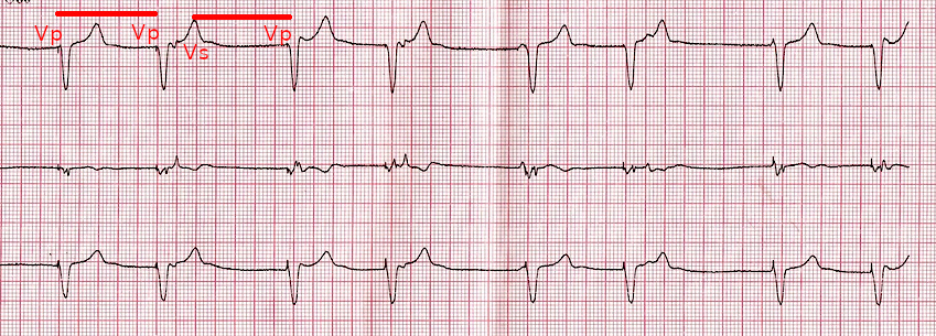
Patient with a pacemaker - Routine FU
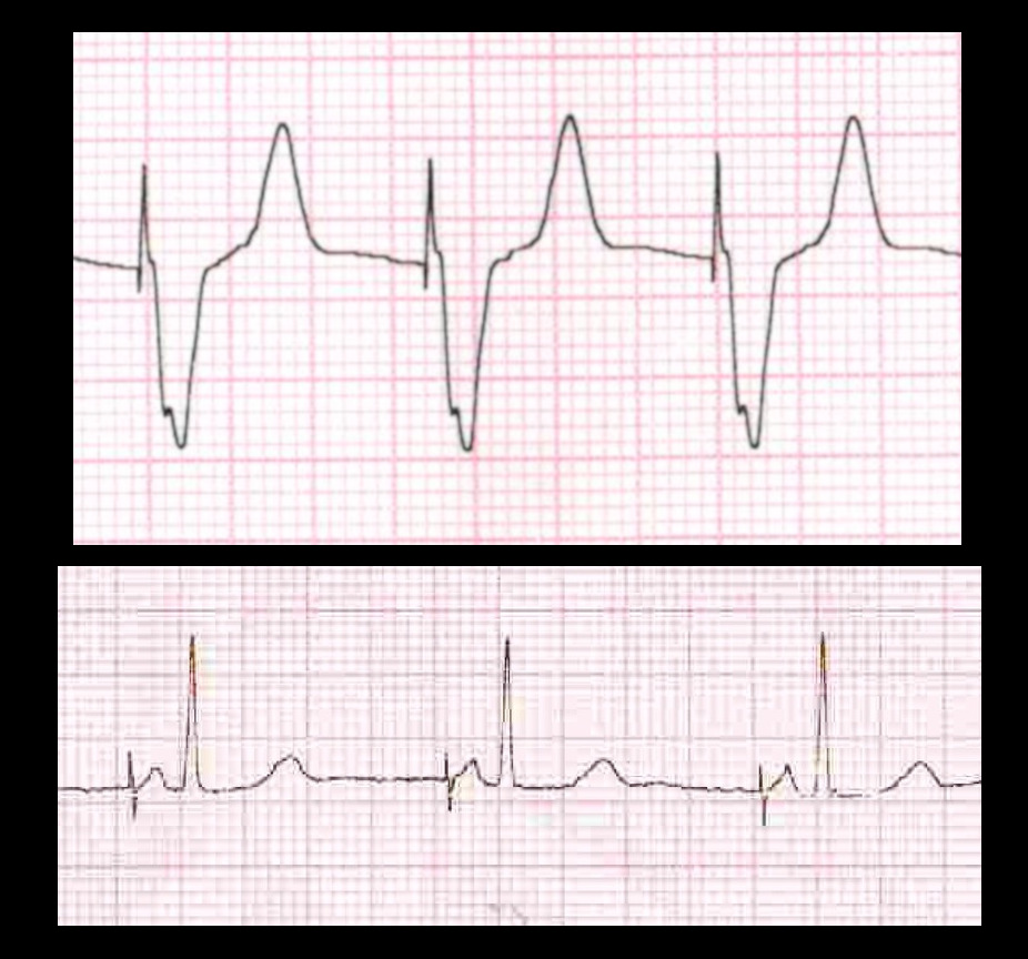
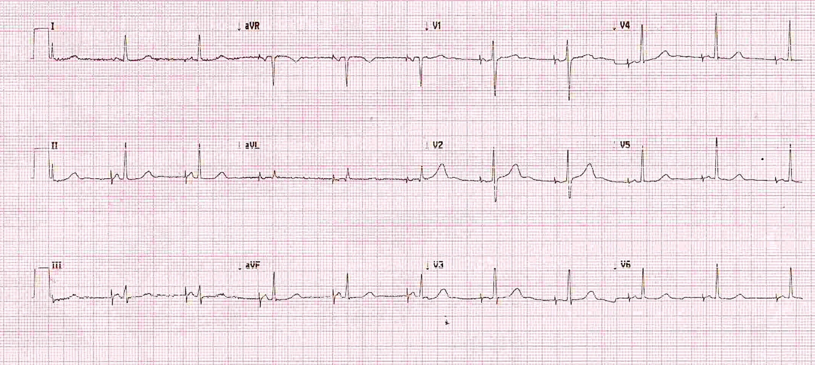
Myth: Pacing spikes not seen = Pacemaker not functioning
- Not pacing because intrinsic rate is faster than set lower rate
- Bipolar pacing - small spikes
- Lead malfunction
- Battery depletion
- Concern if rate is slow and patient is symptomatic
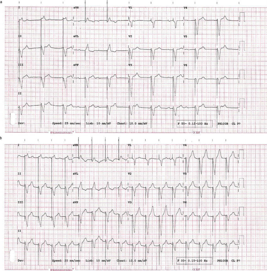
Middle aged female with pacemaker
Elderly male with a pacemaker
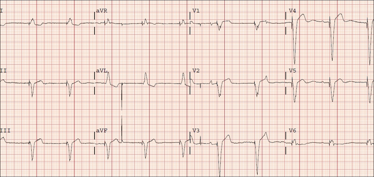
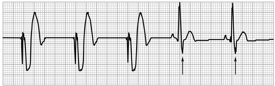
Paced morphology with apical pacing
- LBBB pattern with left axis deviation
- Usually qS in V1, sometimes R wave seen
- Can happen with LV pacing / septal perforation
- But more often occurs in presence of normal location
Pacemaker implanted for complete heart block
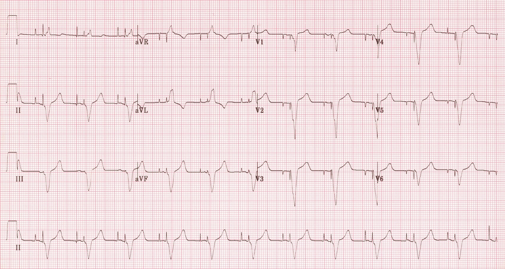
Pacing location - RVOT

BiV CRT
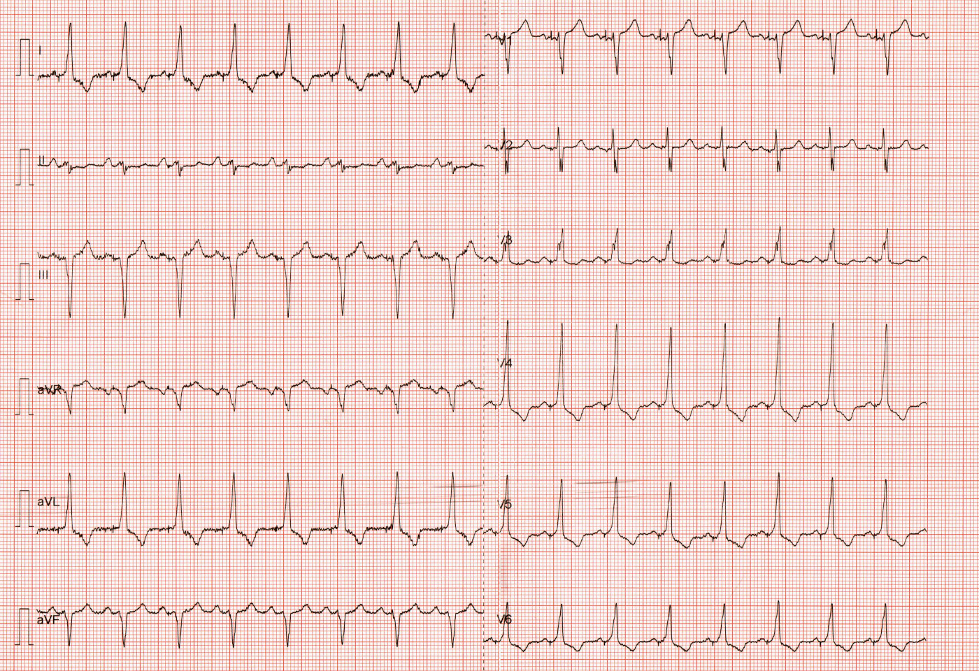
Conduction system pacing
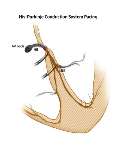
His bundle pacing
Left bundle area pacing
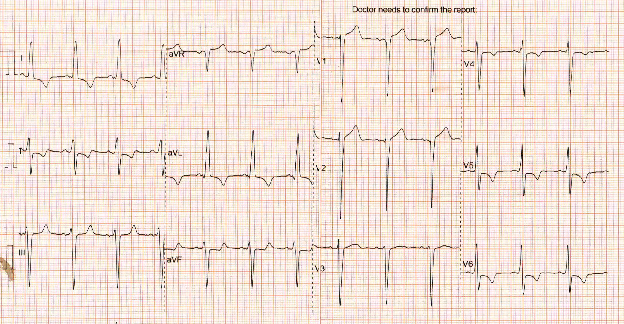
Left bundle area pacing
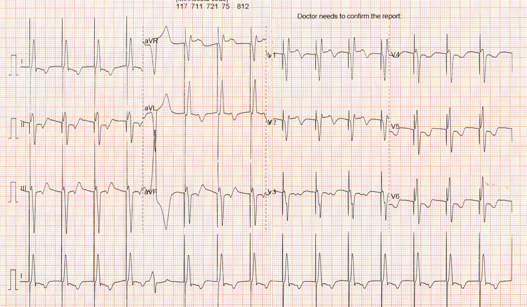
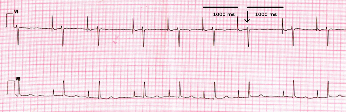
Regular follow up
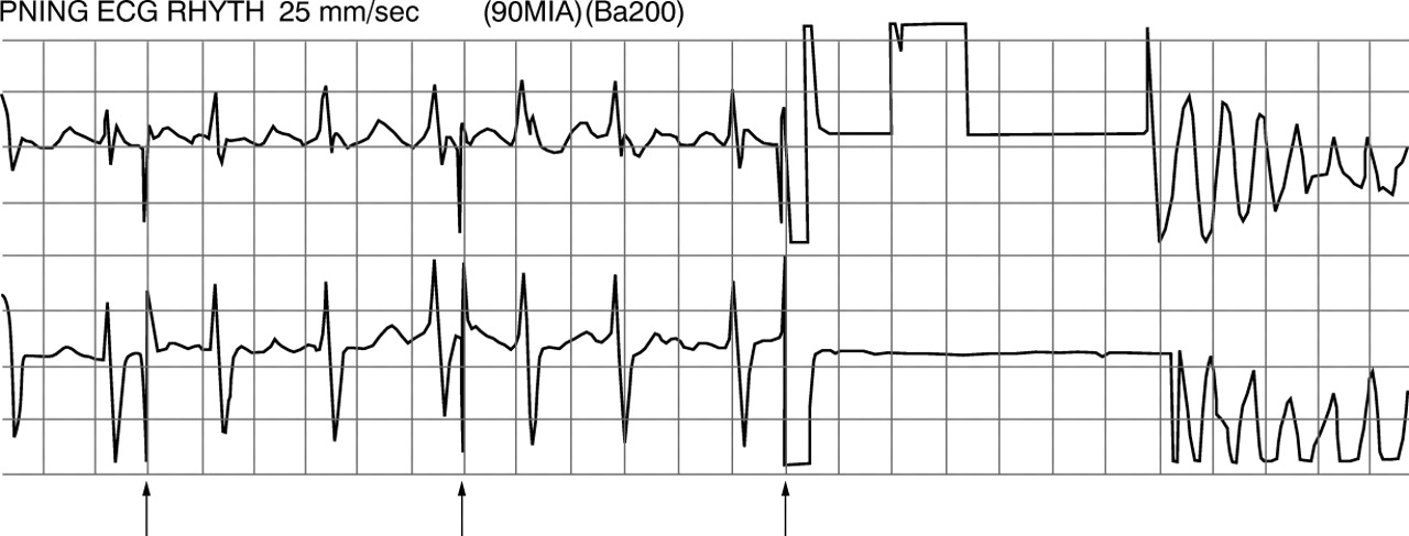
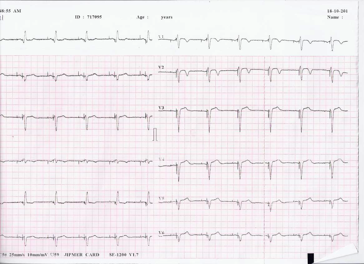
Irregular rhythm
Irregular rhythm explained
AAI with oversensing
Pacemaker implanted 2 weeks back, recurrent syncope
Magnet
Summary
- Understanding basic functioning of pacemaker helps interpret ECGs
- Stepwise approach
- Rate and rhythm
- Identify device
- For each location - Pacing, sensing
- Evaluate rhythm as if there is no pacemaker
- As in other situations, interpret in context of overall clinical findings